

477
16 Fourier Transform Infrared (Ft-Ir) Spectroscopic Imaging …
and subjective process. Efficiency, cost and accuracy can be improved. most importantly, the methods for most tissues are at least several decades old and have
not kept pace with rapid scientific advances that have enabled modern science in a
number of fields. the same morphologic measures also help to diagnose the stage
of disease or other specific characteristics that provide critical input for determining appropriate therapy. It is well recognized that many diagnoses in tissues are not
absolutely accurate and differences in diagnoses between different pathologists as
well as mistakes are often made. hence, the entire process of diagnosing disease and
recommending therapy can be updated with other information. Lately, this process
has been extensively supplemented with molecularly-specific stains. In some cases,
the staining characteristics directly inform molecular medicine to be administered
to the patient. For example, estrogen receptor positive (ER+) breast tumors often
respond to tamoxifen. In general, modern trends in pathology include digital recording and analysis as well as increased integration of molecular content. these two
trends are highly resonant with the goals of using IR imaging for histopathology.
16.2.3 Molecular Histopathology Techniques
At times, definitive diagnoses might not be possible and research questions may
not be answerable by analyzing generic stains. the reasons can be several—the
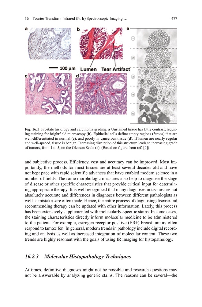
Fig. 16.1 Prostate histology and carcinoma grading. a unstained tissue has little contrast, requiring staining for brightfield microscopy (b). Epithelial cells define empty regions ( lumen) that are
well-differentiated in normal (c), and poorly in cancerous tissue (d). If lumen are nearly regular
and well-spaced, tissue is benign. Increasing disruption of this structure leads to increasing grade
of tumors, from 1 to 5, on the gleason Scale (e). (Based on figure from ref. [2])
16 Fourier Transform Infrared (Ft-Ir) Spectroscopic Imaging …
and subjective process. Efficiency, cost and accuracy can be improved. most importantly, the methods for most tissues are at least several decades old and have
not kept pace with rapid scientific advances that have enabled modern science in a
number of fields. the same morphologic measures also help to diagnose the stage
of disease or other specific characteristics that provide critical input for determining appropriate therapy. It is well recognized that many diagnoses in tissues are not
absolutely accurate and differences in diagnoses between different pathologists as
well as mistakes are often made. hence, the entire process of diagnosing disease and
recommending therapy can be updated with other information. Lately, this process
has been extensively supplemented with molecularly-specific stains. In some cases,
the staining characteristics directly inform molecular medicine to be administered
to the patient. For example, estrogen receptor positive (ER+) breast tumors often
respond to tamoxifen. In general, modern trends in pathology include digital recording and analysis as well as increased integration of molecular content. these two
trends are highly resonant with the goals of using IR imaging for histopathology.
16.2.3 Molecular Histopathology Techniques
At times, definitive diagnoses might not be possible and research questions may
not be answerable by analyzing generic stains. the reasons can be several—the
Fig. 16.1 Prostate histology and carcinoma grading. a unstained tissue has little contrast, requiring staining for brightfield microscopy (b). Epithelial cells define empty regions ( lumen) that are
well-differentiated in normal (c), and poorly in cancerous tissue (d). If lumen are nearly regular
and well-spaced, tissue is benign. Increasing disruption of this structure leads to increasing grade
of tumors, from 1 to 5, on the gleason Scale (e). (Based on figure from ref. [2])