

411
14 Quantitative Analysis of disease Biomarkers using …
analytes in solution and offer several advantages over traditional gold and planar
SERS substrates, such as the ability to localize and concentrate analytes for detection. the LoB platform was used for a SERS-based assay of the cholera toxin.
Figure 14.8 shows a schematic representation of the SERS-based sandwich assay
using gold nanoparticle-coated buoyant silica microspheres.
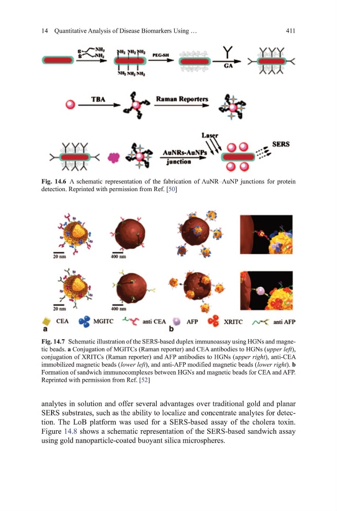
Fig. 14.7 Schematic illustration of the SERS-based duplex immunoassay using hgNs and magnetic beads. a Conjugation of mgItCs (Raman reporter) and CEA antibodies to hgNs ( upper left),
conjugation of XRItCs (Raman reporter) and AFP antibodies to hgNs ( upper right), anti-CEA
immobilized magnetic beads ( lower left), and anti-AFP modified magnetic beads ( lower right). b
Formation of sandwich immunocomplexes between hgNs and magnetic beads for CEA and AFP.
Reprinted with permission from Ref. [52]
Fig. 14.6 A schematic representation of the fabrication of AuNR–AuNP junctions for protein
detection. Reprinted with permission from Ref. [50]
14 Quantitative Analysis of disease Biomarkers using …
analytes in solution and offer several advantages over traditional gold and planar
SERS substrates, such as the ability to localize and concentrate analytes for detection. the LoB platform was used for a SERS-based assay of the cholera toxin.
Figure 14.8 shows a schematic representation of the SERS-based sandwich assay
using gold nanoparticle-coated buoyant silica microspheres.
Fig. 14.7 Schematic illustration of the SERS-based duplex immunoassay using hgNs and magnetic beads. a Conjugation of mgItCs (Raman reporter) and CEA antibodies to hgNs ( upper left),
conjugation of XRItCs (Raman reporter) and AFP antibodies to hgNs ( upper right), anti-CEA
immobilized magnetic beads ( lower left), and anti-AFP modified magnetic beads ( lower right). b
Formation of sandwich immunocomplexes between hgNs and magnetic beads for CEA and AFP.
Reprinted with permission from Ref. [52]
Fig. 14.6 A schematic representation of the fabrication of AuNR–AuNP junctions for protein
detection. Reprinted with permission from Ref. [50]