

406
h. Chon and J. Choo
Z. tian and co-workers [45] fabricated a monolayer SERS substrate of gold
nanoparticles coated with an ultrathin shell of silica (Au/Sio 2 ). this substrate was
used instead of the Au tip traditionally applied in tip-enhanced Raman spectroscopy
(tERS). gold nanoparticles were first chemically synthesized with a core diameter
of 55 nm, onto which ultrathin silica shells were coated to form Au/Sio 2 nanoparticles. Each nanoparticle acts as an Au tip in the tip-enhanced Raman system, and
this technique utilizes thousands of tERS tips to the substrate surface to be probed.
the main advantages of this SERS substrate are its high detection sensitivity and
vast practical applications to various SERS analyses. Figure 14.3 displays the shellisolated nanoparticle monolayer and its working principle.
14.4 SERS-Based Immunoassay Using Magnetic Beads
the SERS-based immunoassay using gold substrates mentioned above has several
limitations. First, an extended incubation time was required for each binding step
because the restriction on molecular diffusion near the surface slows the kinetics of
protein-protein assays. Second, the repetition of washing steps to remove nonspecific binding proteins is inconvenient. third, all of the immune-reagent components
should be immobilized to a surface of a solid substrate in air. however, in many cases, the exposure of proteins to air seriously reduces their biological activity. Finally,
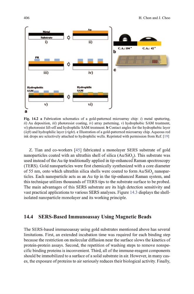
Fig. 14.2 a Fabrication schematics of a gold-patterned microarray chip: i) metal sputtering,
ii) Au deposition, iii) photoresist coating, iv) array patterning, v) hydrophobic SAm treatment,
vi) photoresist lift-off and hydrophilic SAm treatment. b Contact angles for the hydrophobic layer
( left) and hydrophilic layer ( right). c Illustration of a gold-patterned microarray chip. Aqueous red
ink drops are selectively attached to hydrophilic wells. Reprinted with permission from Ref. [19]
h. Chon and J. Choo
Z. tian and co-workers [45] fabricated a monolayer SERS substrate of gold
nanoparticles coated with an ultrathin shell of silica (Au/Sio 2 ). this substrate was
used instead of the Au tip traditionally applied in tip-enhanced Raman spectroscopy
(tERS). gold nanoparticles were first chemically synthesized with a core diameter
of 55 nm, onto which ultrathin silica shells were coated to form Au/Sio 2 nanoparticles. Each nanoparticle acts as an Au tip in the tip-enhanced Raman system, and
this technique utilizes thousands of tERS tips to the substrate surface to be probed.
the main advantages of this SERS substrate are its high detection sensitivity and
vast practical applications to various SERS analyses. Figure 14.3 displays the shellisolated nanoparticle monolayer and its working principle.
14.4 SERS-Based Immunoassay Using Magnetic Beads
the SERS-based immunoassay using gold substrates mentioned above has several
limitations. First, an extended incubation time was required for each binding step
because the restriction on molecular diffusion near the surface slows the kinetics of
protein-protein assays. Second, the repetition of washing steps to remove nonspecific binding proteins is inconvenient. third, all of the immune-reagent components
should be immobilized to a surface of a solid substrate in air. however, in many cases, the exposure of proteins to air seriously reduces their biological activity. Finally,
Fig. 14.2 a Fabrication schematics of a gold-patterned microarray chip: i) metal sputtering,
ii) Au deposition, iii) photoresist coating, iv) array patterning, v) hydrophobic SAm treatment,
vi) photoresist lift-off and hydrophilic SAm treatment. b Contact angles for the hydrophobic layer
( left) and hydrophilic layer ( right). c Illustration of a gold-patterned microarray chip. Aqueous red
ink drops are selectively attached to hydrophilic wells. Reprinted with permission from Ref. [19]