

407
14 Quantitative Analysis of disease Biomarkers using …
in the immobilization-based SERS chip, it is very difficult to fabricate surfaces with
highly reproducible enhancements. As a result, reproducible SERS signals could
not be obtained with different laser spots.
to resolve these problems, a quick and reproducible SERS-based immunoanalysis technique using magnetic beads has been developed. this method does not use
an immobilization procedure on a planar substrate; instead, it uses magnetic beads
as antibody-supporting materials. In this system, the sandwich immunocomplexes
are immobilized to the microtube wall using a small magnetic bar. this technique
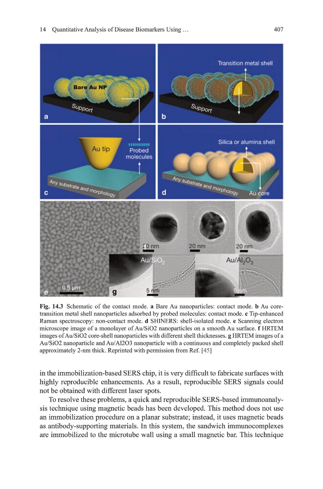
Fig. 14.3 Schematic of the contact mode. a Bare Au nanoparticles: contact mode. b Au coretransition metal shell nanoparticles adsorbed by probed molecules: contact mode. c tip-enhanced
Raman spectroscopy: non-contact mode. d ShINERS: shell-isolated mode. e Scanning electron
microscope image of a monolayer of Au/Sio2 nanoparticles on a smooth Au surface. f hRtEm
images of Au/Sio2 core-shell nanoparticles with different shell thicknesses. g hRtEm images of a
Au/Sio2 nanoparticle and Au/Al2o3 nanoparticle with a continuous and completely packed shell
approximately 2-nm thick. Reprinted with permission from Ref. [45]
14 Quantitative Analysis of disease Biomarkers using …
in the immobilization-based SERS chip, it is very difficult to fabricate surfaces with
highly reproducible enhancements. As a result, reproducible SERS signals could
not be obtained with different laser spots.
to resolve these problems, a quick and reproducible SERS-based immunoanalysis technique using magnetic beads has been developed. this method does not use
an immobilization procedure on a planar substrate; instead, it uses magnetic beads
as antibody-supporting materials. In this system, the sandwich immunocomplexes
are immobilized to the microtube wall using a small magnetic bar. this technique
Fig. 14.3 Schematic of the contact mode. a Bare Au nanoparticles: contact mode. b Au coretransition metal shell nanoparticles adsorbed by probed molecules: contact mode. c tip-enhanced
Raman spectroscopy: non-contact mode. d ShINERS: shell-isolated mode. e Scanning electron
microscope image of a monolayer of Au/Sio2 nanoparticles on a smooth Au surface. f hRtEm
images of Au/Sio2 core-shell nanoparticles with different shell thicknesses. g hRtEm images of a
Au/Sio2 nanoparticle and Au/Al2o3 nanoparticle with a continuous and completely packed shell
approximately 2-nm thick. Reprinted with permission from Ref. [45]