

5 α-Amino Acids In Water: A Review of VCD and ROA Spectra
133
5.5.2.8 L-valine
O
OH
NH 2
C
H 3
CH 3
Scheme 11 (S)-2-amino -3-methylbutanoic acid
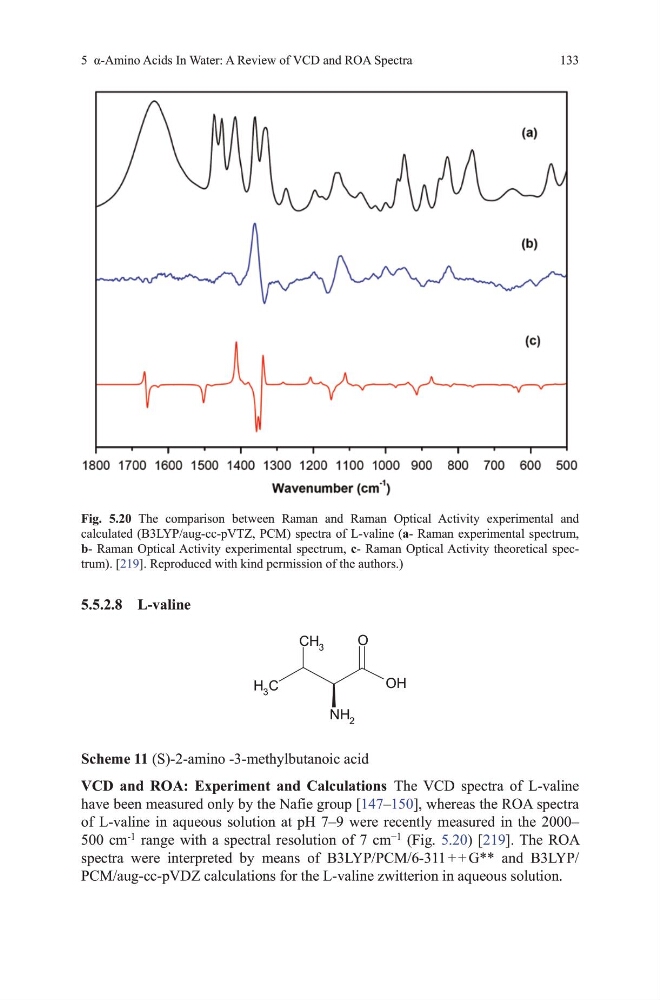
VCD and ROA: Experiment and Calculations the vCd spectra of L-valine
have been measured only by the Nafie group [147–150], whereas the RoA spectra
of L-valine in aqueous solution at ph 7–9 were recently measured in the 2000–
500 cm
-1
range with a spectral resolution of 7 cm
−1
(Fig. 5.20) [219]. the RoA
spectra were interpreted by means of B3LYP/PCm/6-311 + + g** and B3LYP/
PCm/aug-cc-pvdZ calculations for the L-valine zwitterion in aqueous solution.
Fig. 5.20 the comparison between Raman and Raman optical Activity experimental and
calculated (B3LYP/aug-cc-pvtZ, PCm) spectra of L-valine (a- Raman experimental spectrum,
b- Raman optical Activity experimental spectrum, c- Raman optical Activity theoretical spectrum). [219]. Reproduced with kind permission of the authors.)
133
5.5.2.8 L-valine
O
OH
NH 2
C
H 3
CH 3
Scheme 11 (S)-2-amino -3-methylbutanoic acid
VCD and ROA: Experiment and Calculations the vCd spectra of L-valine
have been measured only by the Nafie group [147–150], whereas the RoA spectra
of L-valine in aqueous solution at ph 7–9 were recently measured in the 2000–
500 cm
-1
range with a spectral resolution of 7 cm
−1
(Fig. 5.20) [219]. the RoA
spectra were interpreted by means of B3LYP/PCm/6-311 + + g** and B3LYP/
PCm/aug-cc-pvdZ calculations for the L-valine zwitterion in aqueous solution.
Fig. 5.20 the comparison between Raman and Raman optical Activity experimental and
calculated (B3LYP/aug-cc-pvtZ, PCm) spectra of L-valine (a- Raman experimental spectrum,
b- Raman optical Activity experimental spectrum, c- Raman optical Activity theoretical spectrum). [219]. Reproduced with kind permission of the authors.)