

J. C. Dobrowolsk et al.
134
5.5.2.9 L-phenylalanine, L-tryptophan, L-tyrosine
O
OH
NH 2
O
OH
NH 2
NH
O
OH
NH 2
O
H
(S)-2-amino-3-phenyl
propanoic acid
(2S)-2-amino-3-(1H-indol-3yl)propanoic acid
(S)-2-Amino-3-(4-hydroxyphenyl) propanoic acid
Scheme 12 L-phenylalanine, L-tryptophan, L-tyrosine
VCD and ROA: Experiment and Calculations As for many other amino acids, for
L-phenylalanine, the only vCd measurements were done for the C*h band stretching
and bending vibration bands by the Nafie group in the 1980s [147–150], and in the
form of a film with α-cyclodextrin by Zhang and Polavarapu [38]. For L-tryptophan,
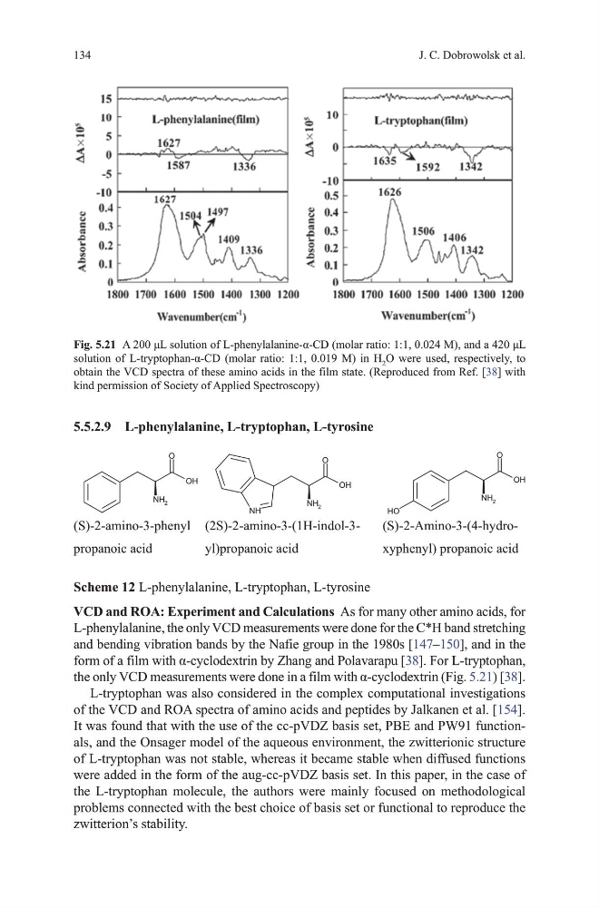
the only VCD measurements were done in a film with α-cyclodextrin (Fig. 5.21) [38].
L-tryptophan was also considered in the complex computational investigations
of the vCd and RoA spectra of amino acids and peptides by Jalkanen et al. [154].
It was found that with the use of the cc-pvdZ basis set, PBE and PW91 functionals, and the onsager model of the aqueous environment, the zwitterionic structure
of L-tryptophan was not stable, whereas it became stable when diffused functions
were added in the form of the aug-cc-pvdZ basis set. In this paper, in the case of
the L-tryptophan molecule, the authors were mainly focused on methodological
problems connected with the best choice of basis set or functional to reproduce the
zwitterion’s stability.
Fig. 5.21 A 200 μL solution of L-phenylalanine-α-CD (molar ratio: 1:1, 0.024 M), and a 420 μL
solution of L-tryptophan-α-CD (molar ratio: 1:1, 0.019 M) in H 2 o were used, respectively, to
obtain the vCd spectra of these amino acids in the film state. (Reproduced from Ref. [38] with
kind permission of Society of Applied Spectroscopy)
134
5.5.2.9 L-phenylalanine, L-tryptophan, L-tyrosine
O
OH
NH 2
O
OH
NH 2
NH
O
OH
NH 2
O
H
(S)-2-amino-3-phenyl
propanoic acid
(2S)-2-amino-3-(1H-indol-3yl)propanoic acid
(S)-2-Amino-3-(4-hydroxyphenyl) propanoic acid
Scheme 12 L-phenylalanine, L-tryptophan, L-tyrosine
VCD and ROA: Experiment and Calculations As for many other amino acids, for
L-phenylalanine, the only vCd measurements were done for the C*h band stretching
and bending vibration bands by the Nafie group in the 1980s [147–150], and in the
form of a film with α-cyclodextrin by Zhang and Polavarapu [38]. For L-tryptophan,
the only VCD measurements were done in a film with α-cyclodextrin (Fig. 5.21) [38].
L-tryptophan was also considered in the complex computational investigations
of the vCd and RoA spectra of amino acids and peptides by Jalkanen et al. [154].
It was found that with the use of the cc-pvdZ basis set, PBE and PW91 functionals, and the onsager model of the aqueous environment, the zwitterionic structure
of L-tryptophan was not stable, whereas it became stable when diffused functions
were added in the form of the aug-cc-pvdZ basis set. In this paper, in the case of
the L-tryptophan molecule, the authors were mainly focused on methodological
problems connected with the best choice of basis set or functional to reproduce the
zwitterion’s stability.
Fig. 5.21 A 200 μL solution of L-phenylalanine-α-CD (molar ratio: 1:1, 0.024 M), and a 420 μL
solution of L-tryptophan-α-CD (molar ratio: 1:1, 0.019 M) in H 2 o were used, respectively, to
obtain the vCd spectra of these amino acids in the film state. (Reproduced from Ref. [38] with
kind permission of Society of Applied Spectroscopy)