

J. C. Dobrowolsk et al.
126
Let us mention that the vCd, IR, and uv-vis spectra of N-acetyl-L-cysteine,
a drug used in detoxifying free radicals, were also measured in aqueous solution
(d 2 o) at several ph levels [137]. the spectra were interpreted by means of B3LYP/6-311 + + g** calculations and molecular dynamics simulations. As found previously for unsubstituted L-cysteine [137], the energetic order of the protonated,
neutral, mono- and doubly-deprotonated N-acetyl-L-cysteine conformers was noticeably changed after the inclusion of the polarisable continuum model. to properly account for the discrepancies noted between the experimental and simulated
vCd spectra, the PCm and the explicit solvent model (preceded by the md simulations of N-acetyl-L-cysteine water clusters) were used. the IR and vCd intensities
computed for N-acetyl-L-cysteine clusters with three or four water molecules at
the B3LYP/6-311 + + g** level with and without the PCm indicated that combination of explicit and implicit solvation models provided better agreement between
theory and experiment, allowing for finding the conformational distributions of
N-acetyl-L-cysteine in water and hydrogen bonding interactions between the solute
and water molecules.
5.5.2.4 L-serine
O
OH
NH 2
O
H
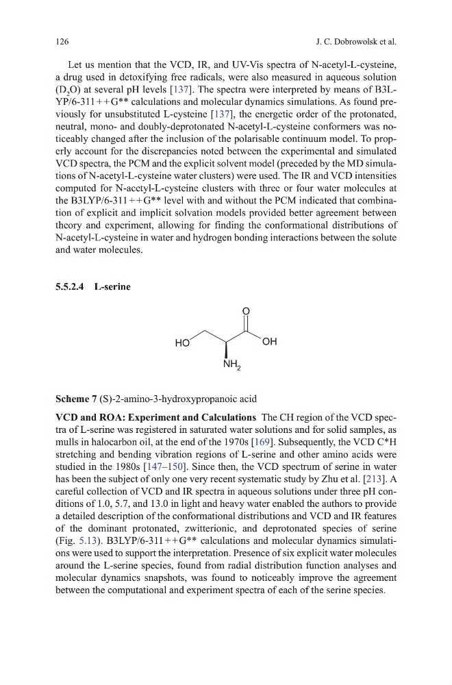
Scheme 7 (S)-2-amino-3-hydroxypropanoic acid
VCD and ROA: Experiment and Calculations the Ch region of the vCd spectra of L-serine was registered in saturated water solutions and for solid samples, as
mulls in halocarbon oil, at the end of the 1970s [169]. Subsequently, the vCd C*h
stretching and bending vibration regions of L-serine and other amino acids were
studied in the 1980s [147–150]. Since then, the vCd spectrum of serine in water
has been the subject of only one very recent systematic study by Zhu et al. [213]. A
careful collection of vCd and IR spectra in aqueous solutions under three ph conditions of 1.0, 5.7, and 13.0 in light and heavy water enabled the authors to provide
a detailed description of the conformational distributions and vCd and IR features
of the dominant protonated, zwitterionic, and deprotonated species of serine
(Fig. 5.13). B3LYP/6-311 + + g** calculations and molecular dynamics simulations were used to support the interpretation. Presence of six explicit water molecules
around the L-serine species, found from radial distribution function analyses and
molecular dynamics snapshots, was found to noticeably improve the agreement
between the computational and experiment spectra of each of the serine species.
126
Let us mention that the vCd, IR, and uv-vis spectra of N-acetyl-L-cysteine,
a drug used in detoxifying free radicals, were also measured in aqueous solution
(d 2 o) at several ph levels [137]. the spectra were interpreted by means of B3LYP/6-311 + + g** calculations and molecular dynamics simulations. As found previously for unsubstituted L-cysteine [137], the energetic order of the protonated,
neutral, mono- and doubly-deprotonated N-acetyl-L-cysteine conformers was noticeably changed after the inclusion of the polarisable continuum model. to properly account for the discrepancies noted between the experimental and simulated
vCd spectra, the PCm and the explicit solvent model (preceded by the md simulations of N-acetyl-L-cysteine water clusters) were used. the IR and vCd intensities
computed for N-acetyl-L-cysteine clusters with three or four water molecules at
the B3LYP/6-311 + + g** level with and without the PCm indicated that combination of explicit and implicit solvation models provided better agreement between
theory and experiment, allowing for finding the conformational distributions of
N-acetyl-L-cysteine in water and hydrogen bonding interactions between the solute
and water molecules.
5.5.2.4 L-serine
O
OH
NH 2
O
H
Scheme 7 (S)-2-amino-3-hydroxypropanoic acid
VCD and ROA: Experiment and Calculations the Ch region of the vCd spectra of L-serine was registered in saturated water solutions and for solid samples, as
mulls in halocarbon oil, at the end of the 1970s [169]. Subsequently, the vCd C*h
stretching and bending vibration regions of L-serine and other amino acids were
studied in the 1980s [147–150]. Since then, the vCd spectrum of serine in water
has been the subject of only one very recent systematic study by Zhu et al. [213]. A
careful collection of vCd and IR spectra in aqueous solutions under three ph conditions of 1.0, 5.7, and 13.0 in light and heavy water enabled the authors to provide
a detailed description of the conformational distributions and vCd and IR features
of the dominant protonated, zwitterionic, and deprotonated species of serine
(Fig. 5.13). B3LYP/6-311 + + g** calculations and molecular dynamics simulations were used to support the interpretation. Presence of six explicit water molecules
around the L-serine species, found from radial distribution function analyses and
molecular dynamics snapshots, was found to noticeably improve the agreement
between the computational and experiment spectra of each of the serine species.