

5 α-Amino Acids In Water: A Review of VCD and ROA Spectra
125
that the ν(OH) and ν(NH 2 ) and β(oh 2 ) and β(Nh 2 ) ranges of the vCd spectra were
especially useful for discriminating between different chiral forms of intermolecular
hydrogen-bonding complexes. In particular, they characterised the vCd chirality
transfer phenomenon [67, 211, 212] occurring from chiral L-cysteine to an achiral
water molecule after complex formation in which the water molecule acquired significant rotational strengths whose signs changed in line with the geometry of the
complex. They suggested ν
a
(Nh 2 ), β(Nh 2 ), and ν(C = O) to be relatively insensitive
to complex structural changes.
Very recently, Kamiński et al. measured and thoroughly interpreted the VCD,
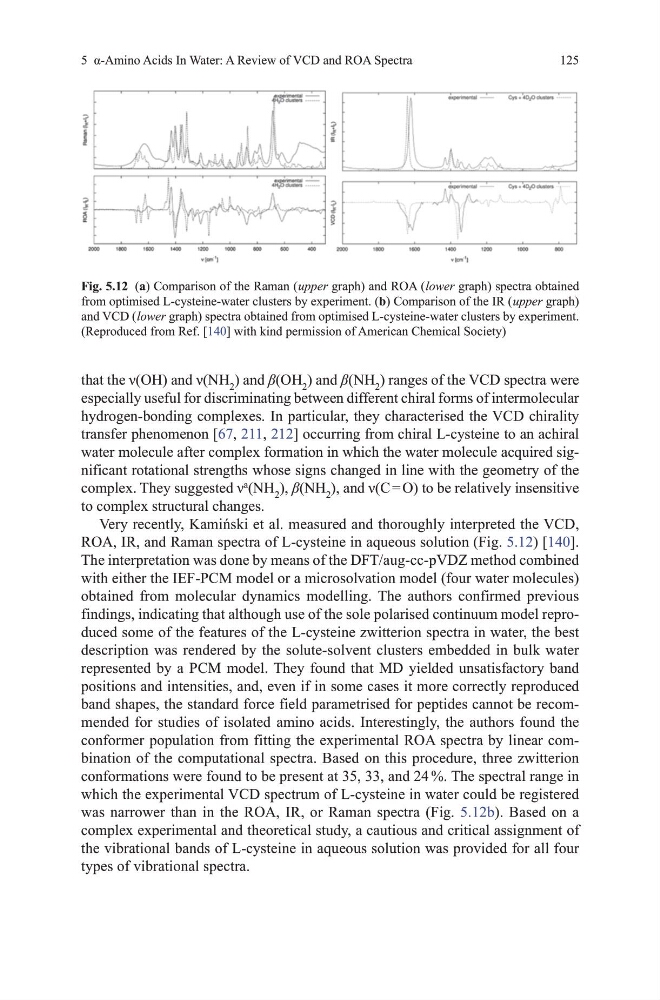
RoA, IR, and Raman spectra of L-cysteine in aqueous solution (Fig. 5.12) [140].
the interpretation was done by means of the dFt/aug-cc-pvdZ method combined
with either the IEF-PCm model or a microsolvation model (four water molecules)
obtained from molecular dynamics modelling. the authors confirmed previous
findings, indicating that although use of the sole polarised continuum model reproduced some of the features of the L-cysteine zwitterion spectra in water, the best
description was rendered by the solute-solvent clusters embedded in bulk water
represented by a PCm model. they found that md yielded unsatisfactory band
positions and intensities, and, even if in some cases it more correctly reproduced
band shapes, the standard force field parametrised for peptides cannot be recommended for studies of isolated amino acids. Interestingly, the authors found the
conformer population from fitting the experimental RoA spectra by linear combination of the computational spectra. Based on this procedure, three zwitterion
conformations were found to be present at 35, 33, and 24 %. the spectral range in
which the experimental vCd spectrum of L-cysteine in water could be registered
was narrower than in the RoA, IR, or Raman spectra (Fig. 5.12b). Based on a
complex experimental and theoretical study, a cautious and critical assignment of
the vibrational bands of L-cysteine in aqueous solution was provided for all four
types of vibrational spectra.
Fig. 5.12 (a) Comparison of the Raman ( upper graph) and RoA ( lower graph) spectra obtained
from optimised L-cysteine-water clusters by experiment. (b) Comparison of the IR ( upper graph)
and vCd ( lower graph) spectra obtained from optimised L-cysteine-water clusters by experiment.
(Reproduced from Ref. [140] with kind permission of American Chemical Society)
125
that the ν(OH) and ν(NH 2 ) and β(oh 2 ) and β(Nh 2 ) ranges of the vCd spectra were
especially useful for discriminating between different chiral forms of intermolecular
hydrogen-bonding complexes. In particular, they characterised the vCd chirality
transfer phenomenon [67, 211, 212] occurring from chiral L-cysteine to an achiral
water molecule after complex formation in which the water molecule acquired significant rotational strengths whose signs changed in line with the geometry of the
complex. They suggested ν
a
(Nh 2 ), β(Nh 2 ), and ν(C = O) to be relatively insensitive
to complex structural changes.
Very recently, Kamiński et al. measured and thoroughly interpreted the VCD,
RoA, IR, and Raman spectra of L-cysteine in aqueous solution (Fig. 5.12) [140].
the interpretation was done by means of the dFt/aug-cc-pvdZ method combined
with either the IEF-PCm model or a microsolvation model (four water molecules)
obtained from molecular dynamics modelling. the authors confirmed previous
findings, indicating that although use of the sole polarised continuum model reproduced some of the features of the L-cysteine zwitterion spectra in water, the best
description was rendered by the solute-solvent clusters embedded in bulk water
represented by a PCm model. they found that md yielded unsatisfactory band
positions and intensities, and, even if in some cases it more correctly reproduced
band shapes, the standard force field parametrised for peptides cannot be recommended for studies of isolated amino acids. Interestingly, the authors found the
conformer population from fitting the experimental RoA spectra by linear combination of the computational spectra. Based on this procedure, three zwitterion
conformations were found to be present at 35, 33, and 24 %. the spectral range in
which the experimental vCd spectrum of L-cysteine in water could be registered
was narrower than in the RoA, IR, or Raman spectra (Fig. 5.12b). Based on a
complex experimental and theoretical study, a cautious and critical assignment of
the vibrational bands of L-cysteine in aqueous solution was provided for all four
types of vibrational spectra.
Fig. 5.12 (a) Comparison of the Raman ( upper graph) and RoA ( lower graph) spectra obtained
from optimised L-cysteine-water clusters by experiment. (b) Comparison of the IR ( upper graph)
and vCd ( lower graph) spectra obtained from optimised L-cysteine-water clusters by experiment.
(Reproduced from Ref. [140] with kind permission of American Chemical Society)