

5 α-Amino Acids In Water: A Review of VCD and ROA Spectra
127
the RoA spectra of neutral L-serine conformers in water were calculated by
Pecul in 2006 [209]. As for cysteine, the main aim of the study was to relate the
changeability of the RoA spectrum with structural variations occurring as the serine
conformation changed. however, unlike for cysteine for which interactions of side
Sh groups are relatively weak, the formation of hydrogen bonds by the side oh
group was found to be the deciding factor determining the RoA intensities of the
side chain vibrations [209].
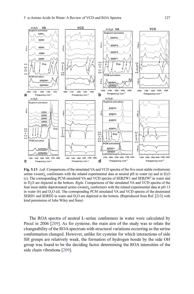
Fig. 5.13 Left. Comparisons of the simulated vA and vCd spectra of the five most stable zwitterionic
serine–(water) 6 conformers with the related experimental data at neutral ph in water (a) and in d 2 o
(c). the corresponding PCm simulated vA and vCd spectra of SERZW1 and SERZW
2
in water and
in d 2 o are depicted at the bottom. Right. Comparisons of the simulated vA and vCd spectra of the
four most stable deprotonated serine–(water) 6 conformers with the related experimental data at ph 13
in water (b) and d 2 o (d). the corresponding PCm simulated vA and vCd spectra of the deuterated
SERd1 and SERd2 in water and d 2 o are depicted at the bottom. (Reproduced from Ref. [213] with
kind permission of John Wiley and Sons)
127
the RoA spectra of neutral L-serine conformers in water were calculated by
Pecul in 2006 [209]. As for cysteine, the main aim of the study was to relate the
changeability of the RoA spectrum with structural variations occurring as the serine
conformation changed. however, unlike for cysteine for which interactions of side
Sh groups are relatively weak, the formation of hydrogen bonds by the side oh
group was found to be the deciding factor determining the RoA intensities of the
side chain vibrations [209].
Fig. 5.13 Left. Comparisons of the simulated vA and vCd spectra of the five most stable zwitterionic
serine–(water) 6 conformers with the related experimental data at neutral ph in water (a) and in d 2 o
(c). the corresponding PCm simulated vA and vCd spectra of SERZW1 and SERZW
2
in water and
in d 2 o are depicted at the bottom. Right. Comparisons of the simulated vA and vCd spectra of the
four most stable deprotonated serine–(water) 6 conformers with the related experimental data at ph 13
in water (b) and d 2 o (d). the corresponding PCm simulated vA and vCd spectra of the deuterated
SERd1 and SERd2 in water and d 2 o are depicted at the bottom. (Reproduced from Ref. [213] with
kind permission of John Wiley and Sons)