

might be somewhat altered compared to that of a ‘true’ one-photon absorption
spectrum. An invaluable support to the experimental results is provided by theoretical simulations of the one-photon absorption profile, as will be discussed below.
However, state-of-the-art action spectroscopy is, beyond no doubt, a highly reliable
tool for unraveling a substantial information about main transitions in one-photon
absorption profiles.
5.3.2 Photo-electron Spectra
The prompt action absorption spectrum obtained at ELISA only indirectly shows
that electron detachment is operative. To investigate this in greater detail we
measured photo-electron spectra of the deprotonated GFP chromophore at several
wavelengths within and beyond the first photo-absorption band. These
measurements give the vertical detachment energy (VDE), and, importantly, they
also reveal a competition between direct and indirect electron emission, which are
alternative ‘action’ routes to internal conversion (see Fig. 5.7). The direct and
indirect electron emission channels have different signatures in the photo-electron
spectra, which make their relative importance evident.
The photoelectron data presented here have been obtained at the SEP-II laboratory [46] at the Department of Physics and Astronomy, Aarhus University. A
nano-second laser is used to create photoelectrons from the deprotonated HBDI
chromophore. The electrons are accelerated under velocity-map imaging conditions
towards a Multi-Channel Plate (MCP) detector (labeled E-MCP in Fig. 5.12), where
their position and time-of-flight are recorded. The main ion beam and neutral
fragments pass through a hole in the center of the E-MCP detector. A deflector is
used to steer away the main ion beam after the electron spectrometer, while the
neutral fragments are counted by a second MCP detector. Thus, the position of
electrons and the time-of flight of both the electrons and neutral molecules are
measured. Photodetachment results in very low energy electrons, which under
normal operating conditions continue co-linearly with the ion beam and exit the
spectrometer region undetectable through the hole in the center of the detector.
Therefore, a small magnetic field has been introduced, which causes the electrons to
be slightly deflected from the central ion-beam axis and hit the detector.
A Monte-Carlo algorithm is used to produce the electron-energy distribution
(Fig. 5.13) from the measured radial distribution [35]. At low photon energy, the
data consists of a low-energy peak, whose shape essentially does not change with
wavelength. This fact clearly indicates that PD here occurs resonantly through
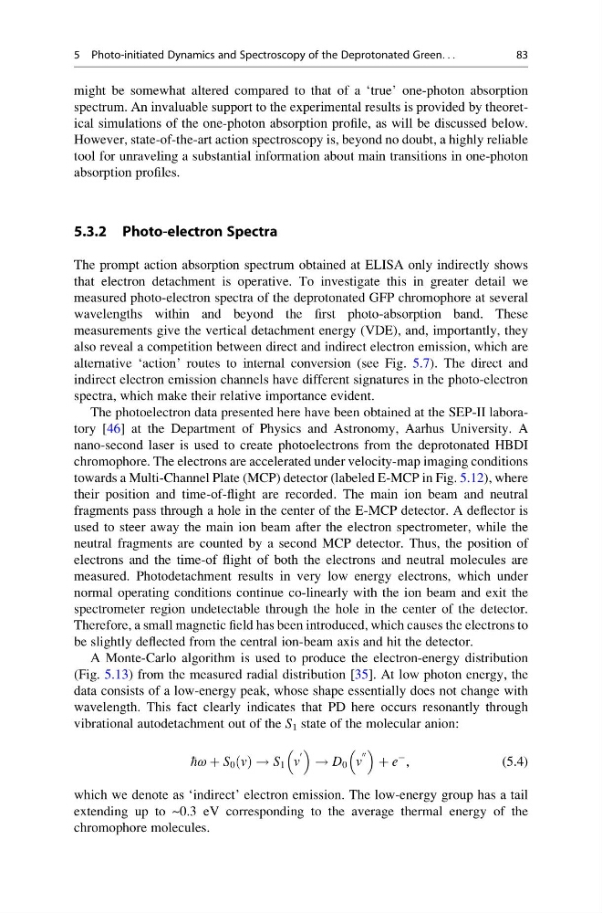
vibrational autodetachment out of the S 1 state of the molecular anion:
hω þ S 0 v
ð Þ ! S 1 v
0
! D 0 v
00
þ e
À ,
(5.4)
which we denote as ‘indirect’ electron emission. The low-energy group has a tail
extending up to ~0.3 eV corresponding to the average thermal energy of the
chromophore molecules.
5 Photo-initiated Dynamics and Spectroscopy of the Deprotonated Green. . .
83
spectrum. An invaluable support to the experimental results is provided by theoretical simulations of the one-photon absorption profile, as will be discussed below.
However, state-of-the-art action spectroscopy is, beyond no doubt, a highly reliable
tool for unraveling a substantial information about main transitions in one-photon
absorption profiles.
5.3.2 Photo-electron Spectra
The prompt action absorption spectrum obtained at ELISA only indirectly shows
that electron detachment is operative. To investigate this in greater detail we
measured photo-electron spectra of the deprotonated GFP chromophore at several
wavelengths within and beyond the first photo-absorption band. These
measurements give the vertical detachment energy (VDE), and, importantly, they
also reveal a competition between direct and indirect electron emission, which are
alternative ‘action’ routes to internal conversion (see Fig. 5.7). The direct and
indirect electron emission channels have different signatures in the photo-electron
spectra, which make their relative importance evident.
The photoelectron data presented here have been obtained at the SEP-II laboratory [46] at the Department of Physics and Astronomy, Aarhus University. A
nano-second laser is used to create photoelectrons from the deprotonated HBDI
chromophore. The electrons are accelerated under velocity-map imaging conditions
towards a Multi-Channel Plate (MCP) detector (labeled E-MCP in Fig. 5.12), where
their position and time-of-flight are recorded. The main ion beam and neutral
fragments pass through a hole in the center of the E-MCP detector. A deflector is
used to steer away the main ion beam after the electron spectrometer, while the
neutral fragments are counted by a second MCP detector. Thus, the position of
electrons and the time-of flight of both the electrons and neutral molecules are
measured. Photodetachment results in very low energy electrons, which under
normal operating conditions continue co-linearly with the ion beam and exit the
spectrometer region undetectable through the hole in the center of the detector.
Therefore, a small magnetic field has been introduced, which causes the electrons to
be slightly deflected from the central ion-beam axis and hit the detector.
A Monte-Carlo algorithm is used to produce the electron-energy distribution
(Fig. 5.13) from the measured radial distribution [35]. At low photon energy, the
data consists of a low-energy peak, whose shape essentially does not change with
wavelength. This fact clearly indicates that PD here occurs resonantly through
vibrational autodetachment out of the S 1 state of the molecular anion:
hω þ S 0 v
ð Þ ! S 1 v
0
! D 0 v
00
þ e
À ,
(5.4)
which we denote as ‘indirect’ electron emission. The low-energy group has a tail
extending up to ~0.3 eV corresponding to the average thermal energy of the
chromophore molecules.
5 Photo-initiated Dynamics and Spectroscopy of the Deprotonated Green. . .
83