

between two bases, there will be two excited-state wavefunctions that can be
approximated by linear combinations of the two wavefunctions from the uncoupled
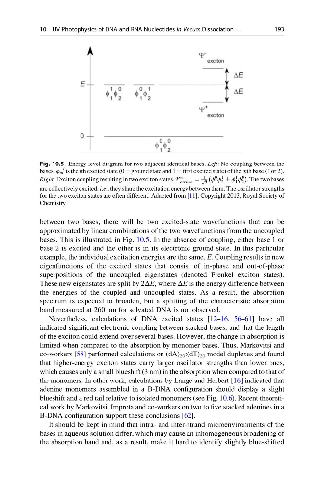
bases. This is illustrated in Fig. 10.5. In the absence of coupling, either base 1 or
base 2 is excited and the other is in its electronic ground state. In this particular
example, the individual excitation energies are the same, E. Coupling results in new
eigenfunctions of the excited states that consist of in-phase and out-of-phase
superpositions of the uncoupled eigenstates (denoted Frenkel exciton states).
These new eigenstates are split by 2ΔE, where ΔE is the energy difference between
the energies of the coupled and uncoupled states. As a result, the absorption
spectrum is expected to broaden, but a splitting of the characteristic absorption
band measured at 260 nm for solvated DNA is not observed.
Nevertheless, calculations of DNA excited states [12–16, 56–61] have all
indicated significant electronic coupling between stacked bases, and that the length
of the exciton could extend over several bases. However, the change in absorption is
limited when compared to the absorption by monomer bases. Thus, Markovitsi and
co-workers [58] performed calculations on (dA) 20 :(dT) 20 model duplexes and found
that higher-energy exciton states carry larger oscillator strengths than lower ones,
which causes only a small blueshift (3 nm) in the absorption when compared to that of
the monomers. In other work, calculations by Lange and Herbert [16] indicated that
adenine monomers assembled in a B-DNA configuration should display a slight
blueshift and a red tail relative to isolated monomers (see Fig. 10.6). Recent theoretical work by Markovitsi, Improta and co-workers on two to five stacked adenines in a
B-DNA configuration support these conclusions [62].
It should be kept in mind that intra- and inter-strand microenvironments of the
bases in aqueous solution differ, which may cause an inhomogeneous broadening of
the absorption band and, as a result, make it hard to identify slightly blue-shifted
Fig. 10.5 Energy level diagram for two adjacent identical bases. Left: No coupling between the
bases. φ m
i is the ith excited state (0 ¼ ground state and 1 ¼ first excited state) of the mth base (1 or 2).
Right: Exciton coupling resulting in two exciton states, Ψ
Æ
exciton ¼
1 ffiffi
2
p ϕ
0
1 ϕ
1
2 Æ ϕ
1
1 ϕ
0
2
À
Á
. The two bases
are collectively excited, i.e., they share the excitation energy between them. The oscillator strengths
for the two exciton states are often different. Adapted from [11]. Copyright 2013, Royal Society of
Chemistry
10 UV Photophysics of DNA and RNA Nucleotides In Vacuo: Dissociation. . .
193
approximated by linear combinations of the two wavefunctions from the uncoupled
bases. This is illustrated in Fig. 10.5. In the absence of coupling, either base 1 or
base 2 is excited and the other is in its electronic ground state. In this particular
example, the individual excitation energies are the same, E. Coupling results in new
eigenfunctions of the excited states that consist of in-phase and out-of-phase
superpositions of the uncoupled eigenstates (denoted Frenkel exciton states).
These new eigenstates are split by 2ΔE, where ΔE is the energy difference between
the energies of the coupled and uncoupled states. As a result, the absorption
spectrum is expected to broaden, but a splitting of the characteristic absorption
band measured at 260 nm for solvated DNA is not observed.
Nevertheless, calculations of DNA excited states [12–16, 56–61] have all
indicated significant electronic coupling between stacked bases, and that the length
of the exciton could extend over several bases. However, the change in absorption is
limited when compared to the absorption by monomer bases. Thus, Markovitsi and
co-workers [58] performed calculations on (dA) 20 :(dT) 20 model duplexes and found
that higher-energy exciton states carry larger oscillator strengths than lower ones,
which causes only a small blueshift (3 nm) in the absorption when compared to that of
the monomers. In other work, calculations by Lange and Herbert [16] indicated that
adenine monomers assembled in a B-DNA configuration should display a slight
blueshift and a red tail relative to isolated monomers (see Fig. 10.6). Recent theoretical work by Markovitsi, Improta and co-workers on two to five stacked adenines in a
B-DNA configuration support these conclusions [62].
It should be kept in mind that intra- and inter-strand microenvironments of the
bases in aqueous solution differ, which may cause an inhomogeneous broadening of
the absorption band and, as a result, make it hard to identify slightly blue-shifted
Fig. 10.5 Energy level diagram for two adjacent identical bases. Left: No coupling between the
bases. φ m
i is the ith excited state (0 ¼ ground state and 1 ¼ first excited state) of the mth base (1 or 2).
Right: Exciton coupling resulting in two exciton states, Ψ
Æ
exciton ¼
1 ffiffi
2
p ϕ
0
1 ϕ
1
2 Æ ϕ
1
1 ϕ
0
2
À
Á
. The two bases
are collectively excited, i.e., they share the excitation energy between them. The oscillator strengths
for the two exciton states are often different. Adapted from [11]. Copyright 2013, Royal Society of
Chemistry
10 UV Photophysics of DNA and RNA Nucleotides In Vacuo: Dissociation. . .
193