

schemes. In line with the observed competition between detachment and relaxation,
Gabelica et al. [24] found that there were two deexcitation pathways following 260nm excitation of DNA multianions, one involving IC and subsequent dissociation and
the other being electron detachment. However, a repulsive Coulomb barrier present
in the multianions can hinder electron detachment (which would require electron
tunnelling through the barrier), causing IC to be a more favourable process. Furthermore, it is also possible that dissociation of the radical anions can occur following
electron detachment. For more discussion on the competition between electron
detachment and internal conversion, we refer to Chap. 5 by Andersen and
Bochenkova.
Finally, we note that, by comparing the time scales for dissociation (tens of μs)
and solvent cooling (ca. 10 ps), one can estimate a quantum yield of ca. 10
À6 for
“thermal” dissociation of nucleotides in the condensed phase, which could still lead
to low levels of DNA photodamage. Of course, it is likely that solvent reorientation
for such large molecules would be the rate-limiting step for condensed-phase
fragmentation, which would slow this process even further, rendering photodamage
by vibrational predissociation an even rarer event.
10.4 Optical Properties of Nucleotides In Vacuo: Action Spectra
The difference in absorption between single bases and oligonucleotides containing
stacked bases is limited [19, 26], which at first seems to indicate that the bases do
not electronically couple in the excited states. In the case of electronic coupling
0
1
2
3
0
100
0.0
2.0x10
3
0.0
2.0x10
3
4.0x10
3
0.0
2.0x10
4
4.0x10
4
Time (ms)
dA
2–
4
dA
–
4
Neutrals counts
dA
–
3
dA
–
2
0
1
2
3
0.0
2.0x10
3
0.0
2.0x10
3
4.0x10
3
0.0
200.0
400.0
0.0
2.0x10
4
4.0x10
4
Time (ms)
dA
2–
4
dA
–
4
Neutrals counts
dA
–
3
dA
–
2
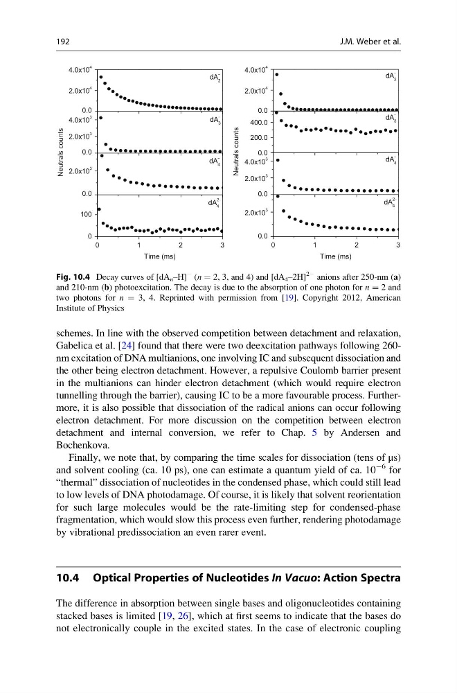
Fig. 10.4 Decay curves of [dA n –H]
À (n ¼ 2, 3, and 4) and [dA 4 –2H]
2À anions after 250-nm (a)
and 210-nm (b) photoexcitation. The decay is due to the absorption of one photon for n ¼ 2 and
two photons for n ¼ 3, 4. Reprinted with permission from [19]. Copyright 2012, American
Institute of Physics
192
J.M. Weber et al.
Gabelica et al. [24] found that there were two deexcitation pathways following 260nm excitation of DNA multianions, one involving IC and subsequent dissociation and
the other being electron detachment. However, a repulsive Coulomb barrier present
in the multianions can hinder electron detachment (which would require electron
tunnelling through the barrier), causing IC to be a more favourable process. Furthermore, it is also possible that dissociation of the radical anions can occur following
electron detachment. For more discussion on the competition between electron
detachment and internal conversion, we refer to Chap. 5 by Andersen and
Bochenkova.
Finally, we note that, by comparing the time scales for dissociation (tens of μs)
and solvent cooling (ca. 10 ps), one can estimate a quantum yield of ca. 10
À6 for
“thermal” dissociation of nucleotides in the condensed phase, which could still lead
to low levels of DNA photodamage. Of course, it is likely that solvent reorientation
for such large molecules would be the rate-limiting step for condensed-phase
fragmentation, which would slow this process even further, rendering photodamage
by vibrational predissociation an even rarer event.
10.4 Optical Properties of Nucleotides In Vacuo: Action Spectra
The difference in absorption between single bases and oligonucleotides containing
stacked bases is limited [19, 26], which at first seems to indicate that the bases do
not electronically couple in the excited states. In the case of electronic coupling
0
1
2
3
0
100
0.0
2.0x10
3
0.0
2.0x10
3
4.0x10
3
0.0
2.0x10
4
4.0x10
4
Time (ms)
dA
2–
4
dA
–
4
Neutrals counts
dA
–
3
dA
–
2
0
1
2
3
0.0
2.0x10
3
0.0
2.0x10
3
4.0x10
3
0.0
200.0
400.0
0.0
2.0x10
4
4.0x10
4
Time (ms)
dA
2–
4
dA
–
4
Neutrals counts
dA
–
3
dA
–
2
Fig. 10.4 Decay curves of [dA n –H]
À (n ¼ 2, 3, and 4) and [dA 4 –2H]
2À anions after 250-nm (a)
and 210-nm (b) photoexcitation. The decay is due to the absorption of one photon for n ¼ 2 and
two photons for n ¼ 3, 4. Reprinted with permission from [19]. Copyright 2012, American
Institute of Physics
192
J.M. Weber et al.