

The question that comes to mind is that of the sensitivity of these action spectra
to the intrinsic properties of the protein ions and to environmental factors. In order
to address the question of the influence of the 3D organisation of the protein on its
UV spectrum, the photodetachment yield spectra of myoglobin proteins
electrosprayed from different solution conditions were recorded. Figure 8.5
presents the UV photodetachment yield of myoglobin in its native form (charge
state 7À) and in its denatured form (charge state 11À). In its native form, the
protein has a heme group at the centre around which the remaining apoprotein folds.
In its denatured form, the apoprotein (i.e., without the heme group) is observed.
1000
1500
2000
0
250
500
m/z
[M-2H]
-
[M-2H]
2225
250
275
300
325
Absorbance (Arb. Unit)
laser wavelength (nm)
225
250
275
300
325
Electron Photodetachment yield
(Arb. Unit)
laser wavelength (nm)
a
b
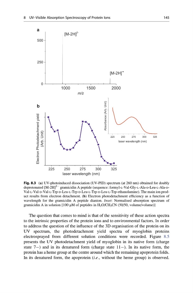
Fig. 8.3 (a) UV-photoinduced dissociation (UV-PID) spectrum (at 260 nm) obtained for doubly
deprotonated [M-2H]
2À gramicidin A peptide (sequence: formyl-L-Val-Gly-L-Ala-D-Leu-L-Ala-DVal-L-Val-D-Val-L-Trp-D-Leu-L-Trp-D-Leu-L-Trp-D-Leu-L-Trp-ethanolamine). The main ion product results from electron detachment. (b) Electron photodetachment efficiency as a function of
wavelength for the gramicidin A peptide dianion. Inset: Normalised absorption spectrum of
gramicidin A in solution [100 μM of peptides in H 2 O/CH 3 CN (50/50, volume/volume)]
8 UV–Visible Absorption Spectroscopy of Protein Ions
145
to the intrinsic properties of the protein ions and to environmental factors. In order
to address the question of the influence of the 3D organisation of the protein on its
UV spectrum, the photodetachment yield spectra of myoglobin proteins
electrosprayed from different solution conditions were recorded. Figure 8.5
presents the UV photodetachment yield of myoglobin in its native form (charge
state 7À) and in its denatured form (charge state 11À). In its native form, the
protein has a heme group at the centre around which the remaining apoprotein folds.
In its denatured form, the apoprotein (i.e., without the heme group) is observed.
1000
1500
2000
0
250
500
m/z
[M-2H]
-
[M-2H]
2225
250
275
300
325
Absorbance (Arb. Unit)
laser wavelength (nm)
225
250
275
300
325
Electron Photodetachment yield
(Arb. Unit)
laser wavelength (nm)
a
b
Fig. 8.3 (a) UV-photoinduced dissociation (UV-PID) spectrum (at 260 nm) obtained for doubly
deprotonated [M-2H]
2À gramicidin A peptide (sequence: formyl-L-Val-Gly-L-Ala-D-Leu-L-Ala-DVal-L-Val-D-Val-L-Trp-D-Leu-L-Trp-D-Leu-L-Trp-D-Leu-L-Trp-ethanolamine). The main ion product results from electron detachment. (b) Electron photodetachment efficiency as a function of
wavelength for the gramicidin A peptide dianion. Inset: Normalised absorption spectrum of
gramicidin A in solution [100 μM of peptides in H 2 O/CH 3 CN (50/50, volume/volume)]
8 UV–Visible Absorption Spectroscopy of Protein Ions
145