

The ππ* band observed for the denatured myoglobin is red-shifted by 10 nm in
comparison to the corresponding native form. Charge states are different, and the
red-shift observed may be explained in part by the increase in negative charges in
the denatured form. Note that in this case, contrary to our insulin example, no
deprotonation of chromophores was expected due to the change in the charge states
(there are 21 acidic residues in myoglobin). Note also that gas-phase spectra are
blue-shifted in comparison to the solution spectrum of myoglobin under native
conditions. The prediction of the shifts due to the solvent results from a subtle
balance between different mechanisms resulting in opposite effects. The theoretical
efforts that are necessary to predict the optical properties of entire proteins with
influence of conformation and solvation are still out of reach.
8.5
Visible Spectroscopy of Protein Anions
Heme proteins, like myoglobin and cytochrome proteins, are metalloproteins
containing a heme prosthetic group, either covalently or noncovalently bound to
the protein. Heme molecules absorb in the visible range. The absorption of hemecontaining moieties in the blue is commonly labelled as the “Soret band”. Protein
chromophores have been extensively studied both in solution and in the gas phase at
the electrostatic heavy-ion storage ring, ELISA, in Aarhus and is discussed in this
volume in Chap. 5 by Andersen and Bochenkova and Chap. 7 by Wyer and
Brøndsted Nielsen [5, 6, 20]. There was evidence that in terms of absorption
there are almost vacuum-like conditions in the hydrophobic interior of certain
proteins like the green fluorescent protein (GFP), as there were no shifts between
the gas-phase spectrum of the bare chromophore and the protein absorption
[M-nH] n1
h
BS
AI
[M-nH]( n-1)-• + e -
RCB
EBE
2
IC
+IVR
h em
n
n
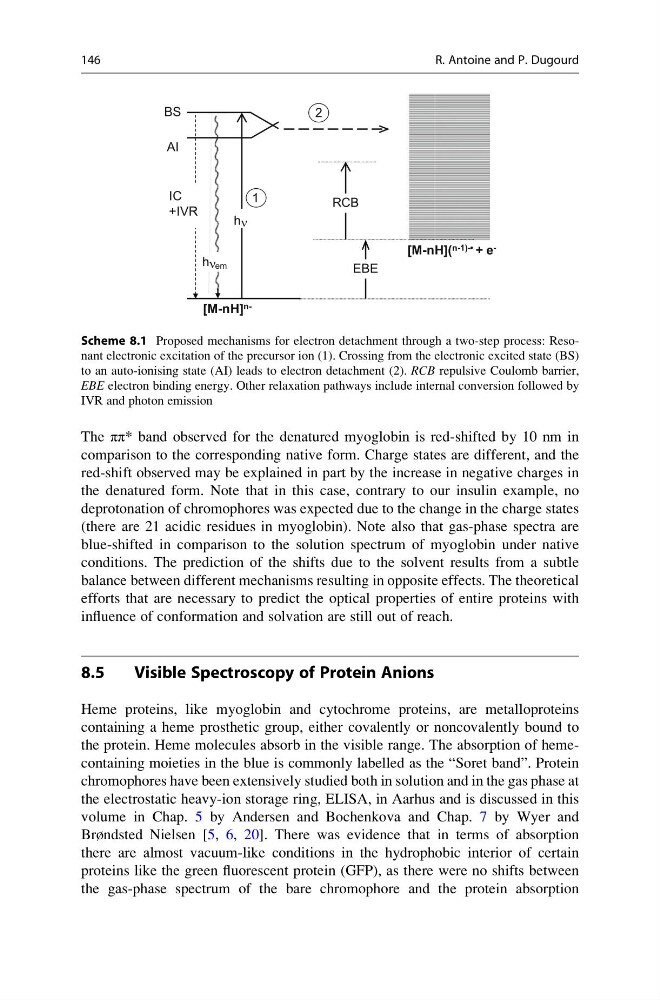
Scheme 8.1 Proposed mechanisms for electron detachment through a two-step process: Resonant electronic excitation of the precursor ion (1). Crossing from the electronic excited state (BS)
to an auto-ionising state (AI) leads to electron detachment (2). RCB repulsive Coulomb barrier,
EBE electron binding energy. Other relaxation pathways include internal conversion followed by
IVR and photon emission
146
R. Antoine and P. Dugourd
comparison to the corresponding native form. Charge states are different, and the
red-shift observed may be explained in part by the increase in negative charges in
the denatured form. Note that in this case, contrary to our insulin example, no
deprotonation of chromophores was expected due to the change in the charge states
(there are 21 acidic residues in myoglobin). Note also that gas-phase spectra are
blue-shifted in comparison to the solution spectrum of myoglobin under native
conditions. The prediction of the shifts due to the solvent results from a subtle
balance between different mechanisms resulting in opposite effects. The theoretical
efforts that are necessary to predict the optical properties of entire proteins with
influence of conformation and solvation are still out of reach.
8.5
Visible Spectroscopy of Protein Anions
Heme proteins, like myoglobin and cytochrome proteins, are metalloproteins
containing a heme prosthetic group, either covalently or noncovalently bound to
the protein. Heme molecules absorb in the visible range. The absorption of hemecontaining moieties in the blue is commonly labelled as the “Soret band”. Protein
chromophores have been extensively studied both in solution and in the gas phase at
the electrostatic heavy-ion storage ring, ELISA, in Aarhus and is discussed in this
volume in Chap. 5 by Andersen and Bochenkova and Chap. 7 by Wyer and
Brøndsted Nielsen [5, 6, 20]. There was evidence that in terms of absorption
there are almost vacuum-like conditions in the hydrophobic interior of certain
proteins like the green fluorescent protein (GFP), as there were no shifts between
the gas-phase spectrum of the bare chromophore and the protein absorption
[M-nH] n1
h
BS
AI
[M-nH]( n-1)-• + e -
RCB
EBE
2
IC
+IVR
h em
n
n
Scheme 8.1 Proposed mechanisms for electron detachment through a two-step process: Resonant electronic excitation of the precursor ion (1). Crossing from the electronic excited state (BS)
to an auto-ionising state (AI) leads to electron detachment (2). RCB repulsive Coulomb barrier,
EBE electron binding energy. Other relaxation pathways include internal conversion followed by
IVR and photon emission
146
R. Antoine and P. Dugourd