

the maximal absorption in the Soret band was found to be at 380 nm which is 20 nm
to the blue of that measured for the bis-pocket siloxyl porphyrin. This suggests that
the cage around the porphyrin is not completely innocent, and that it actually does
perturb the ππ* transition. It should also be mentioned that the peripheral
substituents are different. In the Q-band region increased absorption at ~532 nm
was due to a change in the laser beam profile which resulted in increased overlap
with the ion beam at the second harmonic of the pump laser used to create the
output light. A fit to the data after exclusion of these erroneous points found
maxima at 497 nm and 524 nm for the gaseous ion. This corresponds to an energy
difference of 1,020 cm
À1 . The absorption spectrum of heme
+ in solution where both
axial positions of the heme are taken up by ligands (solvent molecules or anions) is
shown as a dashed blue line in Fig. 7.6a. It displays a broad band between 450 nm
and 550 nm which appears to be formed from two individual band maxima at
500 nm and 539 nm. It is evident that the position of the Q band for isolated 4c ferric
heme, solvated ferric heme, and the cage complex is similar (Fig. 7.6), showing that
in this region the environment does not have a strong effect on the absorption. The
ligand-to-metal transition at high wavelengths is, however, more pronounced in
solution phase revealing a clear band at 626 nm.
In a recent, beautiful experiment, von Helden and co-workers [25] produced
Fe(III)–heme
+ ions in cold helium droplets (16 K). Spectroscopic measurements on
those found the Soret-band position and width to be similar to that measured for the
room-temperature ions, although a slight blue shift (~2 nm) and slight narrowing of
the band (full width half maximum of ~11 nm vs. ~16 nm) at the low-energy side
were seen, in accordance with less hot-band transitions (Fig. 7.9). While a wide
band is expected for room-temperature ions, the still rather broad and unstructured
band for the cold ions was attributed to an ultra-short lifetime in the excited state
(cf., Heisenberg’s uncertainty principle). Indeed, coupling to the S 1 state and d–d
excited states provides an efficient deexcitation route.
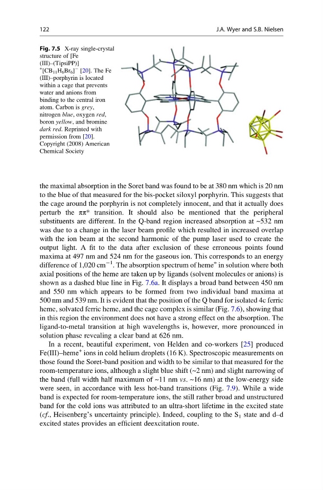
Fig. 7.5 X-ray single-crystal
structure of [Fe
(III)–(TipsiPP)]
+ [CB 11 H 6 Br 6 ]
À [20]. The Fe
(III)–porphyrin is located
within a cage that prevents
water and anions from
binding to the central iron
atom. Carbon is grey,
nitrogen blue, oxygen red,
boron yellow, and bromine
dark red. Reprinted with
permission from [20].
Copyright (2008) American
Chemical Society
122
J.A. Wyer and S.B. Nielsen
to the blue of that measured for the bis-pocket siloxyl porphyrin. This suggests that
the cage around the porphyrin is not completely innocent, and that it actually does
perturb the ππ* transition. It should also be mentioned that the peripheral
substituents are different. In the Q-band region increased absorption at ~532 nm
was due to a change in the laser beam profile which resulted in increased overlap
with the ion beam at the second harmonic of the pump laser used to create the
output light. A fit to the data after exclusion of these erroneous points found
maxima at 497 nm and 524 nm for the gaseous ion. This corresponds to an energy
difference of 1,020 cm
À1 . The absorption spectrum of heme
+ in solution where both
axial positions of the heme are taken up by ligands (solvent molecules or anions) is
shown as a dashed blue line in Fig. 7.6a. It displays a broad band between 450 nm
and 550 nm which appears to be formed from two individual band maxima at
500 nm and 539 nm. It is evident that the position of the Q band for isolated 4c ferric
heme, solvated ferric heme, and the cage complex is similar (Fig. 7.6), showing that
in this region the environment does not have a strong effect on the absorption. The
ligand-to-metal transition at high wavelengths is, however, more pronounced in
solution phase revealing a clear band at 626 nm.
In a recent, beautiful experiment, von Helden and co-workers [25] produced
Fe(III)–heme
+ ions in cold helium droplets (16 K). Spectroscopic measurements on
those found the Soret-band position and width to be similar to that measured for the
room-temperature ions, although a slight blue shift (~2 nm) and slight narrowing of
the band (full width half maximum of ~11 nm vs. ~16 nm) at the low-energy side
were seen, in accordance with less hot-band transitions (Fig. 7.9). While a wide
band is expected for room-temperature ions, the still rather broad and unstructured
band for the cold ions was attributed to an ultra-short lifetime in the excited state
(cf., Heisenberg’s uncertainty principle). Indeed, coupling to the S 1 state and d–d
excited states provides an efficient deexcitation route.
Fig. 7.5 X-ray single-crystal
structure of [Fe
(III)–(TipsiPP)]
+ [CB 11 H 6 Br 6 ]
À [20]. The Fe
(III)–porphyrin is located
within a cage that prevents
water and anions from
binding to the central iron
atom. Carbon is grey,
nitrogen blue, oxygen red,
boron yellow, and bromine
dark red. Reprinted with
permission from [20].
Copyright (2008) American
Chemical Society
122
J.A. Wyer and S.B. Nielsen