

Spectroscopic characterisation of 4c ferric heme in order to obtain a reference
spectrum (or finger-print spectrum) is by conventional methods, however, hampered by the strong affinity of Fe(III) for water and anions. A change in protein
absorption at low pH is therefore difficult to interpret since it may be due to a
different heme microenvironment caused by conformational changes and denaturation, and not necessarily to the formation of 4c Fe(III)–heme. Two very different
approaches were taken to circumvent these obstacles and are described in the
following.
The first measurement of a 4c Fe(III)–porphyrin cation was performed by Fang
et al. [20], who used an elegant synthesis approach to sterically hinder ligation and
produce the 4c [Fe(III)–(TipsiPP)]
+
[CB 11 H 6 Br 6 ]
À (5,10,15,20-tetrakis(2
0 ,6
0 -bis
(triisopropylsiloxy)-phenyl) porphyrinatoiron (III) hexabromocarborane) (see
Fig. 7.5). Maximal absorption in the Soret-band region is at 400 nm, while in the
Q band there is a single band at 505 nm (Fig. 7.6).
An alternative way of recording the absorption spectra of such 4c
Fe(III)–porphyrin ions involves using electrospray ionisation to bring the bare
ions gently and intact into the gas phase. Then isolated ions can be investigated
in vacuo where there are with certainty no external perturbations. This method can
also be used to study more complex ions as axial ligands can be added in a
controlled way. However, a disadvantage with studying ions in the gas phase is
that the number of ions produced is too low to cause a measurable change in the
number of photons in the transmitted light, and conventional absorption spectroscopy cannot be used. This is true even when a trap is used to create a bunch of ions.
Absorption measurements instead rely on the detection of fragmentation or electron
detachment (i.e. action spectroscopy). We note that for iron porphyrins luminescence does not need to be considered as all transitions after photoexcitation are
radiationless; the reason for this is that low-lying d–d excited states of the iron atom
rapidly depopulate the excited state (Fig. 7.7) [24].
The first measurements on the absorption by bare Fe(III)–heme
+ ions (Fig. 7.8)
were performed in vacuo at the ELISA storage ring (see Fig. 7.6b) [21, 22]. There
350
400
450
500
550
600
650
700
0.0
0.5
1.0
1.5
ε × 10
-5
(M
-1
cm
-1
)
Wavelength (nm)
metMb (h)
metMb (h) (NO)
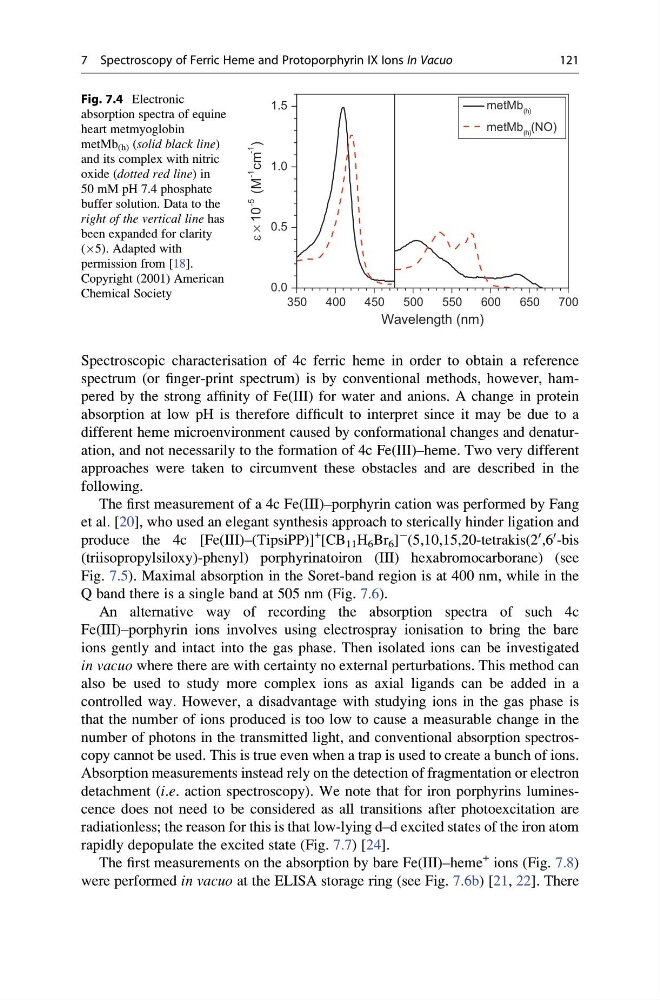
Fig. 7.4 Electronic
absorption spectra of equine
heart metmyoglobin
metMb (h) (solid black line)
and its complex with nitric
oxide (dotted red line) in
50 mM pH 7.4 phosphate
buffer solution. Data to the
right of the vertical line has
been expanded for clarity
(Â5). Adapted with
permission from [18].
Copyright (2001) American
Chemical Society
7 Spectroscopy of Ferric Heme and Protoporphyrin IX Ions In Vacuo
121
spectrum (or finger-print spectrum) is by conventional methods, however, hampered by the strong affinity of Fe(III) for water and anions. A change in protein
absorption at low pH is therefore difficult to interpret since it may be due to a
different heme microenvironment caused by conformational changes and denaturation, and not necessarily to the formation of 4c Fe(III)–heme. Two very different
approaches were taken to circumvent these obstacles and are described in the
following.
The first measurement of a 4c Fe(III)–porphyrin cation was performed by Fang
et al. [20], who used an elegant synthesis approach to sterically hinder ligation and
produce the 4c [Fe(III)–(TipsiPP)]
+
[CB 11 H 6 Br 6 ]
À (5,10,15,20-tetrakis(2
0 ,6
0 -bis
(triisopropylsiloxy)-phenyl) porphyrinatoiron (III) hexabromocarborane) (see
Fig. 7.5). Maximal absorption in the Soret-band region is at 400 nm, while in the
Q band there is a single band at 505 nm (Fig. 7.6).
An alternative way of recording the absorption spectra of such 4c
Fe(III)–porphyrin ions involves using electrospray ionisation to bring the bare
ions gently and intact into the gas phase. Then isolated ions can be investigated
in vacuo where there are with certainty no external perturbations. This method can
also be used to study more complex ions as axial ligands can be added in a
controlled way. However, a disadvantage with studying ions in the gas phase is
that the number of ions produced is too low to cause a measurable change in the
number of photons in the transmitted light, and conventional absorption spectroscopy cannot be used. This is true even when a trap is used to create a bunch of ions.
Absorption measurements instead rely on the detection of fragmentation or electron
detachment (i.e. action spectroscopy). We note that for iron porphyrins luminescence does not need to be considered as all transitions after photoexcitation are
radiationless; the reason for this is that low-lying d–d excited states of the iron atom
rapidly depopulate the excited state (Fig. 7.7) [24].
The first measurements on the absorption by bare Fe(III)–heme
+ ions (Fig. 7.8)
were performed in vacuo at the ELISA storage ring (see Fig. 7.6b) [21, 22]. There
350
400
450
500
550
600
650
700
0.0
0.5
1.0
1.5
ε × 10
-5
(M
-1
cm
-1
)
Wavelength (nm)
metMb (h)
metMb (h) (NO)
Fig. 7.4 Electronic
absorption spectra of equine
heart metmyoglobin
metMb (h) (solid black line)
and its complex with nitric
oxide (dotted red line) in
50 mM pH 7.4 phosphate
buffer solution. Data to the
right of the vertical line has
been expanded for clarity
(Â5). Adapted with
permission from [18].
Copyright (2001) American
Chemical Society
7 Spectroscopy of Ferric Heme and Protoporphyrin IX Ions In Vacuo
121