

22 NIR Imaging
539
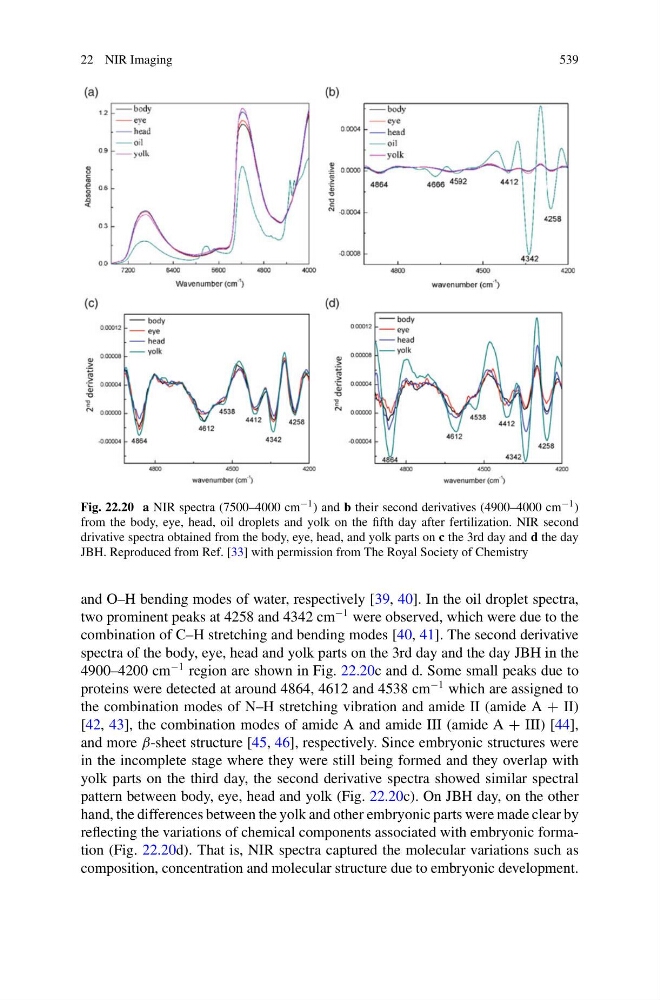
Fig. 22.20 a NIR spectra (7500–4000 cm −1 ) and b their second derivatives (4900–4000 cm −1 )
from the body, eye, head, oil droplets and yolk on the fifth day after fertilization. NIR second
drivative spectra obtained from the body, eye, head, and yolk parts on c the 3rd day and d the day
JBH. Reproduced from Ref. [33] with permission from The Royal Society of Chemistry
and O–H bending modes of water, respectively [39, 40]. In the oil droplet spectra,
two prominent peaks at 4258 and 4342 cm
−1 were observed, which were due to the
combination of C–H stretching and bending modes [40, 41]. The second derivative
spectra of the body, eye, head and yolk parts on the 3rd day and the day JBH in the
4900–4200 cm
−1 region are shown in Fig. 22.20c and d. Some small peaks due to
proteins were detected at around 4864, 4612 and 4538 cm
−1 which are assigned to
the combination modes of N–H stretching vibration and amide II (amide A + II)
[42, 43], the combination modes of amide A and amide III (amide A + III) [44],
and more β-sheet structure [45, 46], respectively. Since embryonic structures were
in the incomplete stage where they were still being formed and they overlap with
yolk parts on the third day, the second derivative spectra showed similar spectral
pattern between body, eye, head and yolk (Fig. 22.20c). On JBH day, on the other
hand, the differences between the yolk and other embryonic parts were made clear by
reflecting the variations of chemical components associated with embryonic formation (Fig. 22.20d). That is, NIR spectra captured the molecular variations such as
composition, concentration and molecular structure due to embryonic development.
539
Fig. 22.20 a NIR spectra (7500–4000 cm −1 ) and b their second derivatives (4900–4000 cm −1 )
from the body, eye, head, oil droplets and yolk on the fifth day after fertilization. NIR second
drivative spectra obtained from the body, eye, head, and yolk parts on c the 3rd day and d the day
JBH. Reproduced from Ref. [33] with permission from The Royal Society of Chemistry
and O–H bending modes of water, respectively [39, 40]. In the oil droplet spectra,
two prominent peaks at 4258 and 4342 cm
−1 were observed, which were due to the
combination of C–H stretching and bending modes [40, 41]. The second derivative
spectra of the body, eye, head and yolk parts on the 3rd day and the day JBH in the
4900–4200 cm
−1 region are shown in Fig. 22.20c and d. Some small peaks due to
proteins were detected at around 4864, 4612 and 4538 cm
−1 which are assigned to
the combination modes of N–H stretching vibration and amide II (amide A + II)
[42, 43], the combination modes of amide A and amide III (amide A + III) [44],
and more β-sheet structure [45, 46], respectively. Since embryonic structures were
in the incomplete stage where they were still being formed and they overlap with
yolk parts on the third day, the second derivative spectra showed similar spectral
pattern between body, eye, head and yolk (Fig. 22.20c). On JBH day, on the other
hand, the differences between the yolk and other embryonic parts were made clear by
reflecting the variations of chemical components associated with embryonic formation (Fig. 22.20d). That is, NIR spectra captured the molecular variations such as
composition, concentration and molecular structure due to embryonic development.