

538
D. Ishikawa et al.
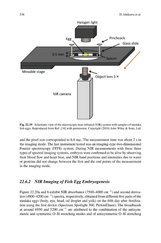
Fig. 22.19 Schematic view of the microscopic near-infrared (NIR) system with samples of medaka
fish eggs. Reproduced from Ref. [34] with permission. Copyright (2018) John Wiley & Sons, Ltd.
and the pixel size corresponded to 6.8 mμ. The measurement time was about 2 s in
the imaging mode. The last instrument tested was an imaging-type two-dimensional
Fourier spectroscopy (ITFS) system. During NIR measurements with these three
types of spectral imaging systems, embryos were confirmed to be alive by observing
their blood flow and heart beat, and NIR band positions and intensities due to water
or proteins did not change between the first and the end points of the measurement
in the imaging mode.
22.6.2 NIR Imaging of Fish Egg Embryogenesis
Figure 22.20a and b exhibit NIR absorbance (7500–4000 cm
−1 ) and second derivative (4900–4200 cm
−1 ) spectra, respectively, obtained from different five parts of the
medaka eggs (body, eye, head, oil droplet and yolk) on the fifth day after fertilization using the first device (Spectrum Spotlight 300, PerkinElmer). The broadbands
at around 6950 and 5200 cm
−1 are attributed to the combination of the antisymmetric and symmetric O–H stretching modes and of antisymmetric O–H stretching
D. Ishikawa et al.
Fig. 22.19 Schematic view of the microscopic near-infrared (NIR) system with samples of medaka
fish eggs. Reproduced from Ref. [34] with permission. Copyright (2018) John Wiley & Sons, Ltd.
and the pixel size corresponded to 6.8 mμ. The measurement time was about 2 s in
the imaging mode. The last instrument tested was an imaging-type two-dimensional
Fourier spectroscopy (ITFS) system. During NIR measurements with these three
types of spectral imaging systems, embryos were confirmed to be alive by observing
their blood flow and heart beat, and NIR band positions and intensities due to water
or proteins did not change between the first and the end points of the measurement
in the imaging mode.
22.6.2 NIR Imaging of Fish Egg Embryogenesis
Figure 22.20a and b exhibit NIR absorbance (7500–4000 cm
−1 ) and second derivative (4900–4200 cm
−1 ) spectra, respectively, obtained from different five parts of the
medaka eggs (body, eye, head, oil droplet and yolk) on the fifth day after fertilization using the first device (Spectrum Spotlight 300, PerkinElmer). The broadbands
at around 6950 and 5200 cm
−1 are attributed to the combination of the antisymmetric and symmetric O–H stretching modes and of antisymmetric O–H stretching