

22 NIR Imaging
537
can reach much deeper in a sample than IR light, NIR imaging can easily investigate
thick samples than IR imaging without much sample preparation.
22.6.1 Application of Three Types of NIR Imaging System
to Biology
Our recent studies investigate the growth of Japanese medaka fish eggs in vivo [32–
36]. They are aimed at exploring embryogenesis at the molecular level by visualizing
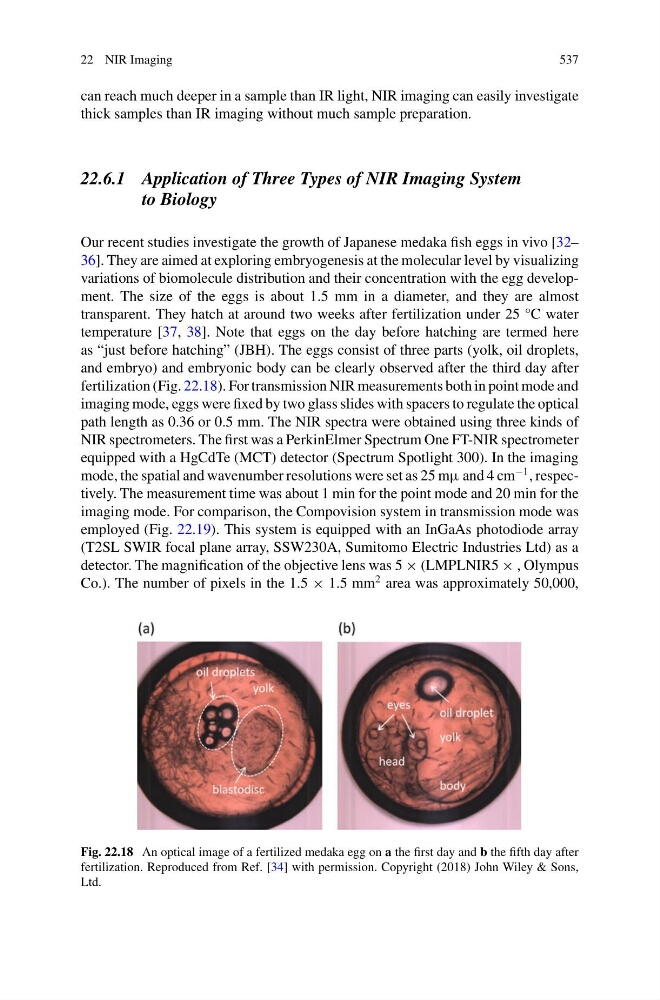
variations of biomolecule distribution and their concentration with the egg development. The size of the eggs is about 1.5 mm in a diameter, and they are almost
transparent. They hatch at around two weeks after fertilization under 25 °C water
temperature [37, 38]. Note that eggs on the day before hatching are termed here
as “just before hatching” (JBH). The eggs consist of three parts (yolk, oil droplets,
and embryo) and embryonic body can be clearly observed after the third day after
fertilization (Fig. 22.18). For transmission NIR measurements both in point mode and
imaging mode, eggs were fixed by two glass slides with spacers to regulate the optical
path length as 0.36 or 0.5 mm. The NIR spectra were obtained using three kinds of
NIR spectrometers. The first was a PerkinElmer Spectrum One FT-NIR spectrometer
equipped with a HgCdTe (MCT) detector (Spectrum Spotlight 300). In the imaging
mode, the spatial and wavenumber resolutions were set as 25 mμ and 4 cm
−1 , respectively. The measurement time was about 1 min for the point mode and 20 min for the
imaging mode. For comparison, the Compovision system in transmission mode was
employed (Fig. 22.19). This system is equipped with an InGaAs photodiode array
(T2SL SWIR focal plane array, SSW230A, Sumitomo Electric Industries Ltd) as a
detector. The magnification of the objective lens was 5 × (LMPLNIR5 × , Olympus
Co.). The number of pixels in the 1.5 × 1.5 mm
2 area was approximately 50,000,
Fig. 22.18 An optical image of a fertilized medaka egg on a the first day and b the fifth day after
fertilization. Reproduced from Ref. [34] with permission. Copyright (2018) John Wiley & Sons,
Ltd.
537
can reach much deeper in a sample than IR light, NIR imaging can easily investigate
thick samples than IR imaging without much sample preparation.
22.6.1 Application of Three Types of NIR Imaging System
to Biology
Our recent studies investigate the growth of Japanese medaka fish eggs in vivo [32–
36]. They are aimed at exploring embryogenesis at the molecular level by visualizing
variations of biomolecule distribution and their concentration with the egg development. The size of the eggs is about 1.5 mm in a diameter, and they are almost
transparent. They hatch at around two weeks after fertilization under 25 °C water
temperature [37, 38]. Note that eggs on the day before hatching are termed here
as “just before hatching” (JBH). The eggs consist of three parts (yolk, oil droplets,
and embryo) and embryonic body can be clearly observed after the third day after
fertilization (Fig. 22.18). For transmission NIR measurements both in point mode and
imaging mode, eggs were fixed by two glass slides with spacers to regulate the optical
path length as 0.36 or 0.5 mm. The NIR spectra were obtained using three kinds of
NIR spectrometers. The first was a PerkinElmer Spectrum One FT-NIR spectrometer
equipped with a HgCdTe (MCT) detector (Spectrum Spotlight 300). In the imaging
mode, the spatial and wavenumber resolutions were set as 25 mμ and 4 cm
−1 , respectively. The measurement time was about 1 min for the point mode and 20 min for the
imaging mode. For comparison, the Compovision system in transmission mode was
employed (Fig. 22.19). This system is equipped with an InGaAs photodiode array
(T2SL SWIR focal plane array, SSW230A, Sumitomo Electric Industries Ltd) as a
detector. The magnification of the objective lens was 5 × (LMPLNIR5 × , Olympus
Co.). The number of pixels in the 1.5 × 1.5 mm
2 area was approximately 50,000,
Fig. 22.18 An optical image of a fertilized medaka egg on a the first day and b the fifth day after
fertilization. Reproduced from Ref. [34] with permission. Copyright (2018) John Wiley & Sons,
Ltd.