

5.13 Analysis of Spectral Characteristics of Radiation Scattered by …
463
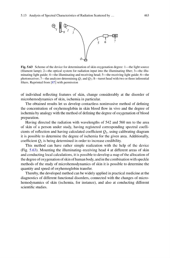
Fig. 5.63 Scheme of the device for determination of skin oxygenation degree: 1—the light source
(filament lamp); 2—the optical system for radiation input into the illuminating fiber; 3—the illuminating light guide; 4—the illuminating and receiving head; 5—the receiving light guide; 6—the
photoreceiver; 7—the analyzer determining Q 1 and Q 2 ; 8—turret head with two or three inferential
filters. Reprinted from [87] with permission
of individual reflecting features of skin, change considerably at the disorder of
microhemodynamics of skin, ischemia in particular.
The obtained results let us develop contactless noninvasive method of defining
the concentration of oxyhemoglobin in skin blood flow in vivo and the degree of
ischemia by analogy with the method of defining the degree of oxygenation of blood
preparation.
Having directed the radiation with wavelengths of 542 and 560 nm to the area
of skin of a person under study, having registered corresponding spectral coefficients of reflection and having calculated coefficient Q 1 , using calibrating diagram
it is possible to determine the degree of ischemia for the given area. Additionally,
coefficient Q 2 is being determined in order to increase credibility.
This method can have rather simple realization with the help of the device
(Fig. 5.63). Mounting the illuminating–receiving head 4 at different areas of skin
and conducting local calculations, it is possible to develop a map of the allocation of
the degree of oxygenation of skin of human body, and in the combination with speckle
methods of the study of microhemodynamics of skin it is possible to determine the
quantity and speed of oxyhemoglobin transfer.
Thereby, the developed method can be widely applied in practical medicine at the
diagnostics of different functional disorders, connected with the changes of microhemodynamics of skin (ischemia, for instance), and also at conducting different
scientific studies.
463
Fig. 5.63 Scheme of the device for determination of skin oxygenation degree: 1—the light source
(filament lamp); 2—the optical system for radiation input into the illuminating fiber; 3—the illuminating light guide; 4—the illuminating and receiving head; 5—the receiving light guide; 6—the
photoreceiver; 7—the analyzer determining Q 1 and Q 2 ; 8—turret head with two or three inferential
filters. Reprinted from [87] with permission
of individual reflecting features of skin, change considerably at the disorder of
microhemodynamics of skin, ischemia in particular.
The obtained results let us develop contactless noninvasive method of defining
the concentration of oxyhemoglobin in skin blood flow in vivo and the degree of
ischemia by analogy with the method of defining the degree of oxygenation of blood
preparation.
Having directed the radiation with wavelengths of 542 and 560 nm to the area
of skin of a person under study, having registered corresponding spectral coefficients of reflection and having calculated coefficient Q 1 , using calibrating diagram
it is possible to determine the degree of ischemia for the given area. Additionally,
coefficient Q 2 is being determined in order to increase credibility.
This method can have rather simple realization with the help of the device
(Fig. 5.63). Mounting the illuminating–receiving head 4 at different areas of skin
and conducting local calculations, it is possible to develop a map of the allocation of
the degree of oxygenation of skin of human body, and in the combination with speckle
methods of the study of microhemodynamics of skin it is possible to determine the
quantity and speed of oxyhemoglobin transfer.
Thereby, the developed method can be widely applied in practical medicine at the
diagnostics of different functional disorders, connected with the changes of microhemodynamics of skin (ischemia, for instance), and also at conducting different
scientific studies.