

158
2 Holographic Microscopy of Phase and Diffuse Objects …
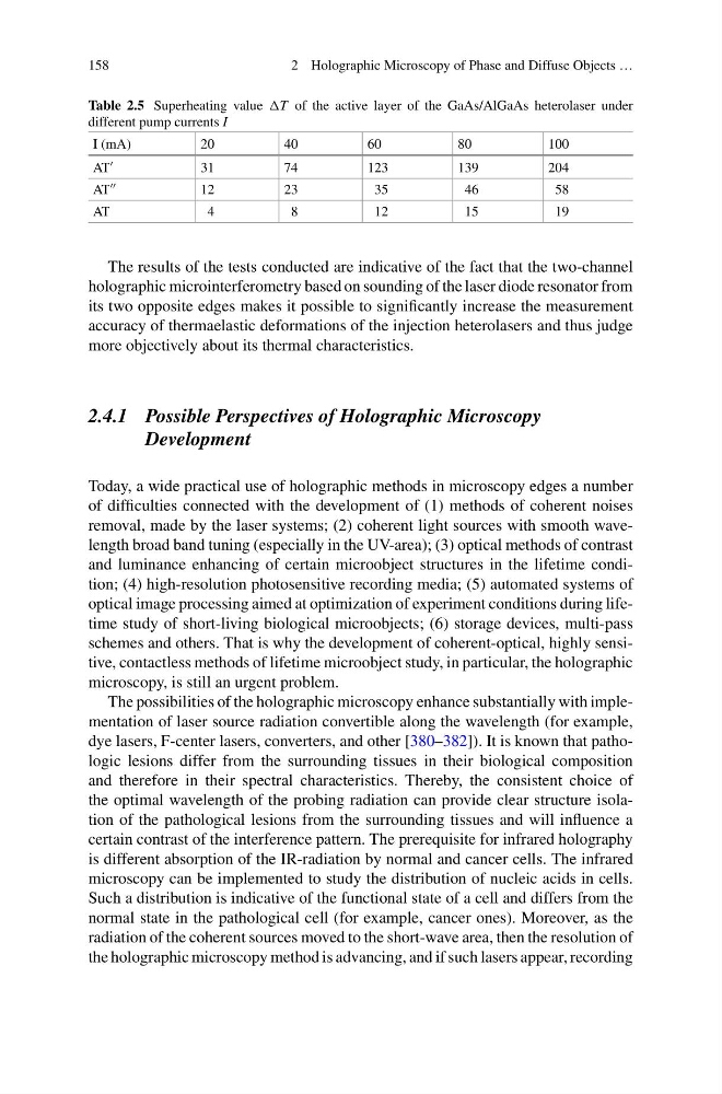
Table 2.5 Superheating value T of the active layer of the GaAs/AlGaAs heterolaser under
different pump currents I
I (mA)
20
40
60
80
100
AT
31
74
123
139
204
AT
12
23
35
46
58
AT
4
8
12
15
19
The results of the tests conducted are indicative of the fact that the two-channel
holographic microinterferometry based on sounding of the laser diode resonator from
its two opposite edges makes it possible to significantly increase the measurement
accuracy of thermaelastic deformations of the injection heterolasers and thus judge
more objectively about its thermal characteristics.
2.4.1 Possible Perspectives of Holographic Microscopy
Development
Today, a wide practical use of holographic methods in microscopy edges a number
of difficulties connected with the development of (1) methods of coherent noises
removal, made by the laser systems; (2) coherent light sources with smooth wavelength broad band tuning (especially in the UV-area); (3) optical methods of contrast
and luminance enhancing of certain microobject structures in the lifetime condition; (4) high-resolution photosensitive recording media; (5) automated systems of
optical image processing aimed at optimization of experiment conditions during lifetime study of short-living biological microobjects; (6) storage devices, multi-pass
schemes and others. That is why the development of coherent-optical, highly sensitive, contactless methods of lifetime microobject study, in particular, the holographic
microscopy, is still an urgent problem.
The possibilities of the holographic microscopy enhance substantially with implementation of laser source radiation convertible along the wavelength (for example,
dye lasers, F-center lasers, converters, and other [380–382]). It is known that pathologic lesions differ from the surrounding tissues in their biological composition
and therefore in their spectral characteristics. Thereby, the consistent choice of
the optimal wavelength of the probing radiation can provide clear structure isolation of the pathological lesions from the surrounding tissues and will influence a
certain contrast of the interference pattern. The prerequisite for infrared holography
is different absorption of the IR-radiation by normal and cancer cells. The infrared
microscopy can be implemented to study the distribution of nucleic acids in cells.
Such a distribution is indicative of the functional state of a cell and differs from the
normal state in the pathological cell (for example, cancer ones). Moreover, as the
radiation of the coherent sources moved to the short-wave area, then the resolution of
the holographic microscopy method is advancing, and if such lasers appear, recording
2 Holographic Microscopy of Phase and Diffuse Objects …
Table 2.5 Superheating value T of the active layer of the GaAs/AlGaAs heterolaser under
different pump currents I
I (mA)
20
40
60
80
100
AT
31
74
123
139
204
AT
12
23
35
46
58
AT
4
8
12
15
19
The results of the tests conducted are indicative of the fact that the two-channel
holographic microinterferometry based on sounding of the laser diode resonator from
its two opposite edges makes it possible to significantly increase the measurement
accuracy of thermaelastic deformations of the injection heterolasers and thus judge
more objectively about its thermal characteristics.
2.4.1 Possible Perspectives of Holographic Microscopy
Development
Today, a wide practical use of holographic methods in microscopy edges a number
of difficulties connected with the development of (1) methods of coherent noises
removal, made by the laser systems; (2) coherent light sources with smooth wavelength broad band tuning (especially in the UV-area); (3) optical methods of contrast
and luminance enhancing of certain microobject structures in the lifetime condition; (4) high-resolution photosensitive recording media; (5) automated systems of
optical image processing aimed at optimization of experiment conditions during lifetime study of short-living biological microobjects; (6) storage devices, multi-pass
schemes and others. That is why the development of coherent-optical, highly sensitive, contactless methods of lifetime microobject study, in particular, the holographic
microscopy, is still an urgent problem.
The possibilities of the holographic microscopy enhance substantially with implementation of laser source radiation convertible along the wavelength (for example,
dye lasers, F-center lasers, converters, and other [380–382]). It is known that pathologic lesions differ from the surrounding tissues in their biological composition
and therefore in their spectral characteristics. Thereby, the consistent choice of
the optimal wavelength of the probing radiation can provide clear structure isolation of the pathological lesions from the surrounding tissues and will influence a
certain contrast of the interference pattern. The prerequisite for infrared holography
is different absorption of the IR-radiation by normal and cancer cells. The infrared
microscopy can be implemented to study the distribution of nucleic acids in cells.
Such a distribution is indicative of the functional state of a cell and differs from the
normal state in the pathological cell (for example, cancer ones). Moreover, as the
radiation of the coherent sources moved to the short-wave area, then the resolution of
the holographic microscopy method is advancing, and if such lasers appear, recording