

2.3 Holographic Study with Electrophysiological Control …
143
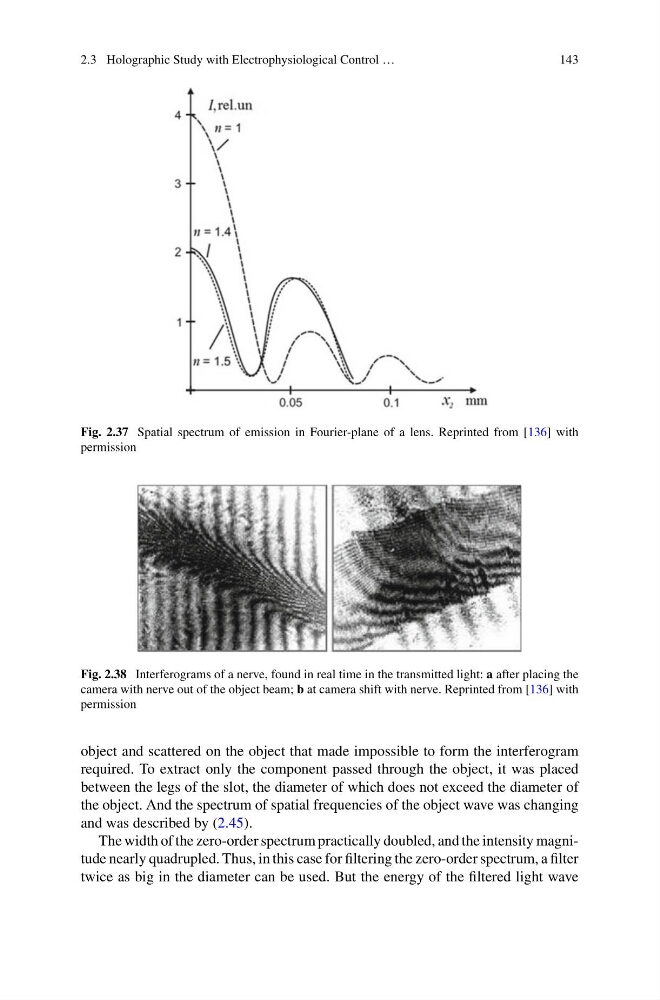
Fig. 2.37 Spatial spectrum of emission in Fourier-plane of a lens. Reprinted from [136] with
permission
Fig. 2.38 Interferograms of a nerve, found in real time in the transmitted light: a after placing the
camera with nerve out of the object beam; b at camera shift with nerve. Reprinted from [136] with
permission
object and scattered on the object that made impossible to form the interferogram
required. To extract only the component passed through the object, it was placed
between the legs of the slot, the diameter of which does not exceed the diameter of
the object. And the spectrum of spatial frequencies of the object wave was changing
and was described by (2.45).
The width of the zero-order spectrum practically doubled, and the intensity magnitude nearly quadrupled. Thus, in this case for filtering the zero-order spectrum, a filter
twice as big in the diameter can be used. But the energy of the filtered light wave
143
Fig. 2.37 Spatial spectrum of emission in Fourier-plane of a lens. Reprinted from [136] with
permission
Fig. 2.38 Interferograms of a nerve, found in real time in the transmitted light: a after placing the
camera with nerve out of the object beam; b at camera shift with nerve. Reprinted from [136] with
permission
object and scattered on the object that made impossible to form the interferogram
required. To extract only the component passed through the object, it was placed
between the legs of the slot, the diameter of which does not exceed the diameter of
the object. And the spectrum of spatial frequencies of the object wave was changing
and was described by (2.45).
The width of the zero-order spectrum practically doubled, and the intensity magnitude nearly quadrupled. Thus, in this case for filtering the zero-order spectrum, a filter
twice as big in the diameter can be used. But the energy of the filtered light wave