

142
2 Holographic Microscopy of Phase and Diffuse Objects …
where R is the radius of the object, n c is the refraction index of the environment; n
is the refraction index of the object.
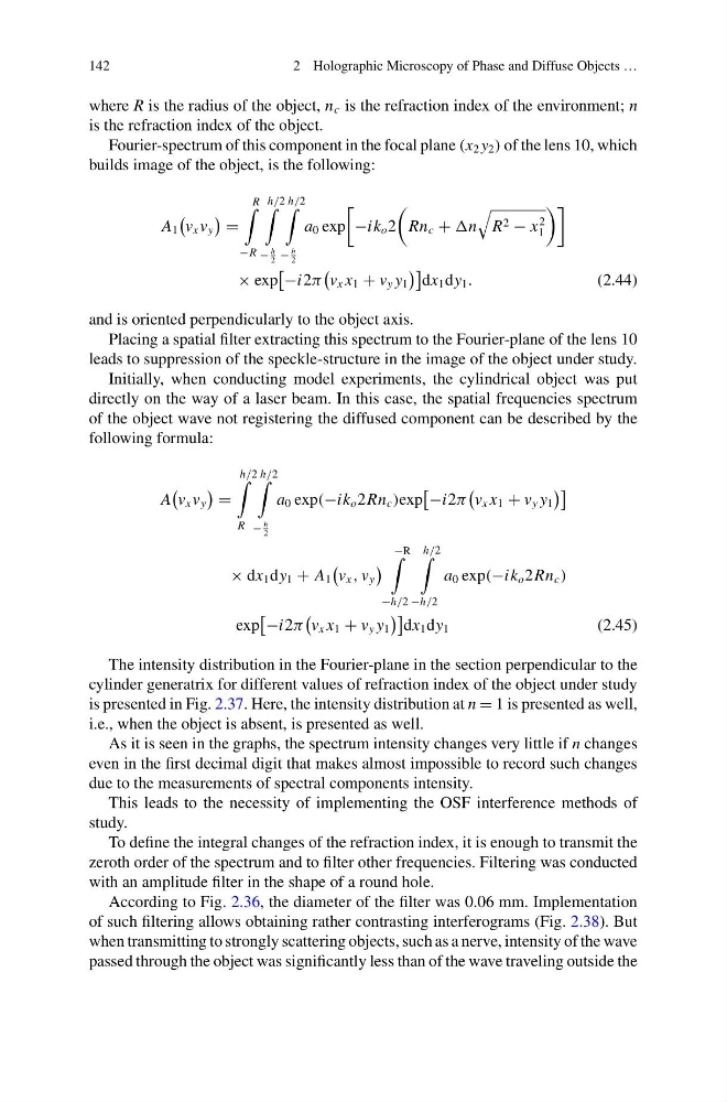
Fourier-spectrum of this component in the focal plane (x 2 y 2 ) of the lens 10, which
builds image of the object, is the following:
A 1
v x v y
=
R
−R
h/2
−
h
2
h/2
−
h
2
a 0 exp
−ik o 2
Rn c + n
R 2 − x
2
1
× exp
−i2π
v x x 1 + v y y 1
dx 1 dy 1 .
(2.44)
and is oriented perpendicularly to the object axis.
Placing a spatial filter extracting this spectrum to the Fourier-plane of the lens 10
leads to suppression of the speckle-structure in the image of the object under study.
Initially, when conducting model experiments, the cylindrical object was put
directly on the way of a laser beam. In this case, the spatial frequencies spectrum
of the object wave not registering the diffused component can be described by the
following formula:
A
v x v y
=
h/2
R
h/2
−
h
2
a 0 exp(−ik o 2Rn c )exp
−i2π
v x x 1 + v y y 1
× dx 1 dy 1 + A 1
v x , v y
−R
−h/2
h/2
−h/2
a 0 exp(−ik o 2Rn c )
exp
−i2π
v x x 1 + v y y 1
dx 1 dy 1
(2.45)
The intensity distribution in the Fourier-plane in the section perpendicular to the
cylinder generatrix for different values of refraction index of the object under study
is presented in Fig. 2.37. Here, the intensity distribution at n = 1 is presented as well,
i.e., when the object is absent, is presented as well.
As it is seen in the graphs, the spectrum intensity changes very little if n changes
even in the first decimal digit that makes almost impossible to record such changes
due to the measurements of spectral components intensity.
This leads to the necessity of implementing the OSF interference methods of
study.
To define the integral changes of the refraction index, it is enough to transmit the
zeroth order of the spectrum and to filter other frequencies. Filtering was conducted
with an amplitude filter in the shape of a round hole.
According to Fig. 2.36, the diameter of the filter was 0.06 mm. Implementation
of such filtering allows obtaining rather contrasting interferograms (Fig. 2.38). But
when transmitting to strongly scattering objects, such as a nerve, intensity of the wave
passed through the object was significantly less than of the wave traveling outside the
2 Holographic Microscopy of Phase and Diffuse Objects …
where R is the radius of the object, n c is the refraction index of the environment; n
is the refraction index of the object.
Fourier-spectrum of this component in the focal plane (x 2 y 2 ) of the lens 10, which
builds image of the object, is the following:
A 1
v x v y
=
R
−R
h/2
−
h
2
h/2
−
h
2
a 0 exp
−ik o 2
Rn c + n
R 2 − x
2
1
× exp
−i2π
v x x 1 + v y y 1
dx 1 dy 1 .
(2.44)
and is oriented perpendicularly to the object axis.
Placing a spatial filter extracting this spectrum to the Fourier-plane of the lens 10
leads to suppression of the speckle-structure in the image of the object under study.
Initially, when conducting model experiments, the cylindrical object was put
directly on the way of a laser beam. In this case, the spatial frequencies spectrum
of the object wave not registering the diffused component can be described by the
following formula:
A
v x v y
=
h/2
R
h/2
−
h
2
a 0 exp(−ik o 2Rn c )exp
−i2π
v x x 1 + v y y 1
× dx 1 dy 1 + A 1
v x , v y
−R
−h/2
h/2
−h/2
a 0 exp(−ik o 2Rn c )
exp
−i2π
v x x 1 + v y y 1
dx 1 dy 1
(2.45)
The intensity distribution in the Fourier-plane in the section perpendicular to the
cylinder generatrix for different values of refraction index of the object under study
is presented in Fig. 2.37. Here, the intensity distribution at n = 1 is presented as well,
i.e., when the object is absent, is presented as well.
As it is seen in the graphs, the spectrum intensity changes very little if n changes
even in the first decimal digit that makes almost impossible to record such changes
due to the measurements of spectral components intensity.
This leads to the necessity of implementing the OSF interference methods of
study.
To define the integral changes of the refraction index, it is enough to transmit the
zeroth order of the spectrum and to filter other frequencies. Filtering was conducted
with an amplitude filter in the shape of a round hole.
According to Fig. 2.36, the diameter of the filter was 0.06 mm. Implementation
of such filtering allows obtaining rather contrasting interferograms (Fig. 2.38). But
when transmitting to strongly scattering objects, such as a nerve, intensity of the wave
passed through the object was significantly less than of the wave traveling outside the