

138
2 Holographic Microscopy of Phase and Diffuse Objects …
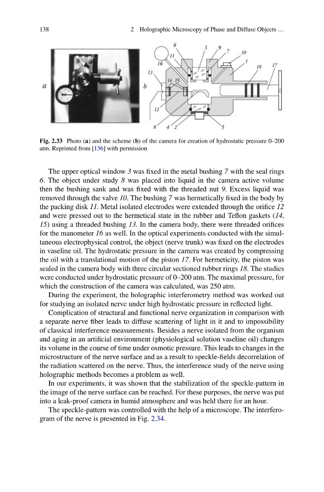
Fig. 2.33 Photo (a) and the scheme (b) of the camera for creation of hydrostatic pressure 0–200
atm. Reprinted from [136] with permission
The upper optical window 3 was fixed in the metal bushing 7 with the seal rings
6. The object under study 8 was placed into liquid in the camera active volume
then the bushing sank and was fixed with the threaded nut 9. Excess liquid was
removed through the valve 10. The bushing 7 was hermetically fixed in the body by
the packing disk 11. Metal isolated electrodes were extended through the orifice 12
and were pressed out to the hermetical state in the rubber and Teflon gaskets (14,
15) using a threaded bushing 13. In the camera body, there were threaded orifices
for the manometer 16 as well. In the optical experiments conducted with the simultaneous electrophysical control, the object (nerve trunk) was fixed on the electrodes
in vaseline oil. The hydrostatic pressure in the camera was created by compressing
the oil with a translational motion of the piston 17. For hermeticity, the piston was
sealed in the camera body with three circular sectioned rubber rings 18. The studies
were conducted under hydrostatic pressure of 0–200 atm. The maximal pressure, for
which the construction of the camera was calculated, was 250 atm.
During the experiment, the holographic interferometry method was worked out
for studying an isolated nerve under high hydrostatic pressure in reflected light.
Complication of structural and functional nerve organization in comparison with
a separate nerve fiber leads to diffuse scattering of light in it and to impossibility
of classical interference measurements. Besides a nerve isolated from the organism
and aging in an artificial environment (physiological solution vaseline oil) changes
its volume in the course of time under osmotic pressure. This leads to changes in the
microstructure of the nerve surface and as a result to speckle-fields decorrelation of
the radiation scattered on the nerve. Thus, the interference study of the nerve using
holographic methods becomes a problem as well.
In our experiments, it was shown that the stabilization of the speckle-pattern in
the image of the nerve surface can be reached. For these purposes, the nerve was put
into a leak-proof camera in humid atmosphere and was held there for an hour.
The speckle-pattern was controlled with the help of a microscope. The interferogram of the nerve is presented in Fig. 2.34.
2 Holographic Microscopy of Phase and Diffuse Objects …
Fig. 2.33 Photo (a) and the scheme (b) of the camera for creation of hydrostatic pressure 0–200
atm. Reprinted from [136] with permission
The upper optical window 3 was fixed in the metal bushing 7 with the seal rings
6. The object under study 8 was placed into liquid in the camera active volume
then the bushing sank and was fixed with the threaded nut 9. Excess liquid was
removed through the valve 10. The bushing 7 was hermetically fixed in the body by
the packing disk 11. Metal isolated electrodes were extended through the orifice 12
and were pressed out to the hermetical state in the rubber and Teflon gaskets (14,
15) using a threaded bushing 13. In the camera body, there were threaded orifices
for the manometer 16 as well. In the optical experiments conducted with the simultaneous electrophysical control, the object (nerve trunk) was fixed on the electrodes
in vaseline oil. The hydrostatic pressure in the camera was created by compressing
the oil with a translational motion of the piston 17. For hermeticity, the piston was
sealed in the camera body with three circular sectioned rubber rings 18. The studies
were conducted under hydrostatic pressure of 0–200 atm. The maximal pressure, for
which the construction of the camera was calculated, was 250 atm.
During the experiment, the holographic interferometry method was worked out
for studying an isolated nerve under high hydrostatic pressure in reflected light.
Complication of structural and functional nerve organization in comparison with
a separate nerve fiber leads to diffuse scattering of light in it and to impossibility
of classical interference measurements. Besides a nerve isolated from the organism
and aging in an artificial environment (physiological solution vaseline oil) changes
its volume in the course of time under osmotic pressure. This leads to changes in the
microstructure of the nerve surface and as a result to speckle-fields decorrelation of
the radiation scattered on the nerve. Thus, the interference study of the nerve using
holographic methods becomes a problem as well.
In our experiments, it was shown that the stabilization of the speckle-pattern in
the image of the nerve surface can be reached. For these purposes, the nerve was put
into a leak-proof camera in humid atmosphere and was held there for an hour.
The speckle-pattern was controlled with the help of a microscope. The interferogram of the nerve is presented in Fig. 2.34.