

2.3 Holographic Study with Electrophysiological Control …
137
Table 2.4 Results of statistical analysis of the electrophysiological study concerning the hyperbary
impact on the isolated nerve
#No. Duration of electrical
stimulation (mcsec)
Analysis data according to the criteria of
Crascal–Wallis, arbit. unit
The level of
criterion
significance
H
M
F
V
V 1
V 2
11
50
3.86 3.86 1.4 × 10 7 1.37 0.56 1.60 <0.0001
100
3.05 3.86 1.1 × 10 7 1.37 0.56 1.60 <0.1
200
3.86 3.86 1.4 × 10 7 1.37 0.56 1.60 <0.0001
500
3.86 3.86 1.4 × 10 7 1.37 0.56 1.60 <0.0001
22
50
4.57 4.57 1.2 × 10 7 2.06 1.31 1.69 <0.0001
100
3.71 4.57 5.60
2.06 1.31 1.69 <0.2
200
3.71 4.57 5.60
2.06 1.31 1.69 <0.2
500
3.71 4.57 5.60
2.06 1.31 1.69 <0.2
Then, the statistics
F =
H (M − k + 1)
(k − 1)(m − H )
,
(2.40)
obeys F-distribution with degrees of freedom
ν 1 = (k − 1)
(k − 1)(M − k + 1) − V
1
2
M V
, ν 2 =
M − k + 1
k − 1
ν 1 .
(2.41)
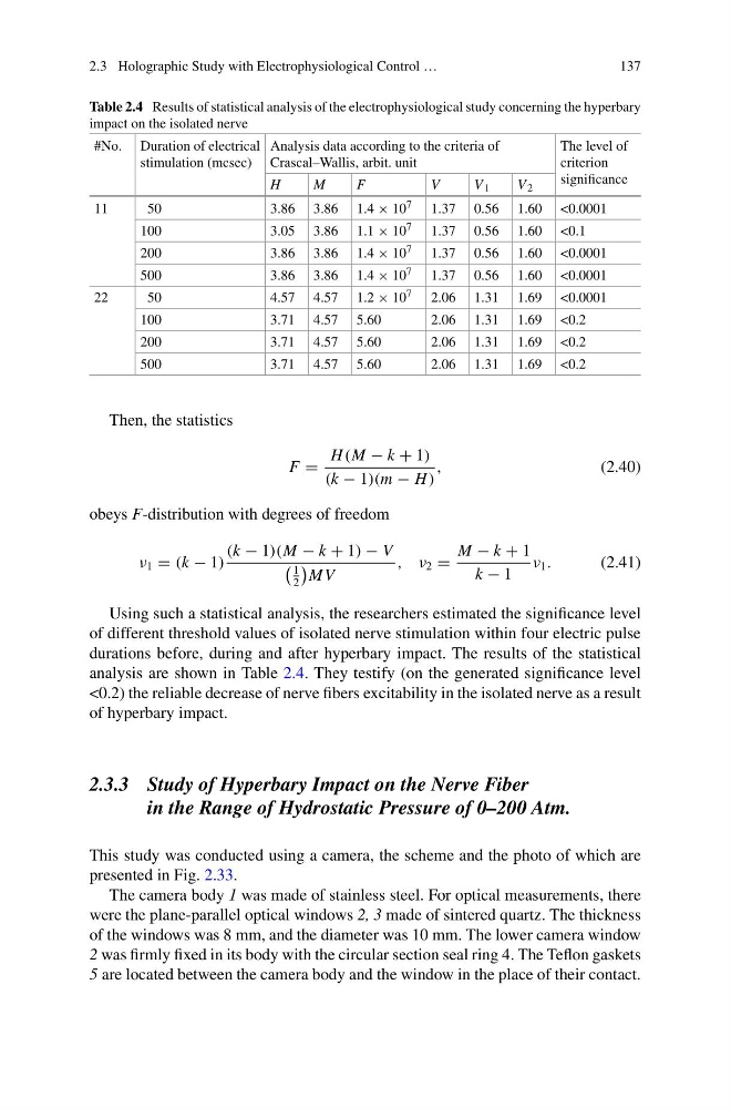
Using such a statistical analysis, the researchers estimated the significance level
of different threshold values of isolated nerve stimulation within four electric pulse
durations before, during and after hyperbary impact. The results of the statistical
analysis are shown in Table 2.4. They testify (on the generated significance level
<0.2) the reliable decrease of nerve fibers excitability in the isolated nerve as a result
of hyperbary impact.
2.3.3 Study of Hyperbary Impact on the Nerve Fiber
in the Range of Hydrostatic Pressure of 0–200 Atm.
This study was conducted using a camera, the scheme and the photo of which are
presented in Fig. 2.33.
The camera body 1 was made of stainless steel. For optical measurements, there
were the plane-parallel optical windows 2, 3 made of sintered quartz. The thickness
of the windows was 8 mm, and the diameter was 10 mm. The lower camera window
2 was firmly fixed in its body with the circular section seal ring 4. The Teflon gaskets
5 are located between the camera body and the window in the place of their contact.
137
Table 2.4 Results of statistical analysis of the electrophysiological study concerning the hyperbary
impact on the isolated nerve
#No. Duration of electrical
stimulation (mcsec)
Analysis data according to the criteria of
Crascal–Wallis, arbit. unit
The level of
criterion
significance
H
M
F
V
V 1
V 2
11
50
3.86 3.86 1.4 × 10 7 1.37 0.56 1.60 <0.0001
100
3.05 3.86 1.1 × 10 7 1.37 0.56 1.60 <0.1
200
3.86 3.86 1.4 × 10 7 1.37 0.56 1.60 <0.0001
500
3.86 3.86 1.4 × 10 7 1.37 0.56 1.60 <0.0001
22
50
4.57 4.57 1.2 × 10 7 2.06 1.31 1.69 <0.0001
100
3.71 4.57 5.60
2.06 1.31 1.69 <0.2
200
3.71 4.57 5.60
2.06 1.31 1.69 <0.2
500
3.71 4.57 5.60
2.06 1.31 1.69 <0.2
Then, the statistics
F =
H (M − k + 1)
(k − 1)(m − H )
,
(2.40)
obeys F-distribution with degrees of freedom
ν 1 = (k − 1)
(k − 1)(M − k + 1) − V
1
2
M V
, ν 2 =
M − k + 1
k − 1
ν 1 .
(2.41)
Using such a statistical analysis, the researchers estimated the significance level
of different threshold values of isolated nerve stimulation within four electric pulse
durations before, during and after hyperbary impact. The results of the statistical
analysis are shown in Table 2.4. They testify (on the generated significance level
<0.2) the reliable decrease of nerve fibers excitability in the isolated nerve as a result
of hyperbary impact.
2.3.3 Study of Hyperbary Impact on the Nerve Fiber
in the Range of Hydrostatic Pressure of 0–200 Atm.
This study was conducted using a camera, the scheme and the photo of which are
presented in Fig. 2.33.
The camera body 1 was made of stainless steel. For optical measurements, there
were the plane-parallel optical windows 2, 3 made of sintered quartz. The thickness
of the windows was 8 mm, and the diameter was 10 mm. The lower camera window
2 was firmly fixed in its body with the circular section seal ring 4. The Teflon gaskets
5 are located between the camera body and the window in the place of their contact.