

2.2 Holographic Study of Structural and Functional Characteristics …
123
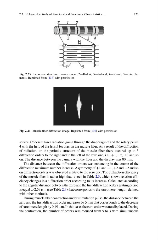
Fig. 2.23 Sarcomere structure: 1—sarcomere; 2—H-disk; 3—A-band; 4—I-band; 5—thin filaments. Reprinted from [136] with permission
Fig. 2.24 Muscle fiber diffraction image. Reprinted from [136] with permission
source. Coherent laser radiation going through the diaphragm 2 and the rotary prism
4 with the help of the lens 5 focuses on the muscle fiber. As a result of the diffraction
of radiation, on the periodic structure of the muscle fiber there occured up to 5
diffraction orders to the right and to the left of the zero one, i.e., +1, ±2, ±3 and so
on. The distance between the camera with the fiber and the display was 80 mm.
The distance between the diffraction orders was enhancing in the course of the
diffraction maximum number increase. Asymmetry of +1 and −1, +2 and −2 and so
on diffraction orders was observed relative to the zero one. The diffraction efficiency
of the muscle fiber is rather high that is seen in Table 2.3, which shows relation efficiency changes in a diffraction order according to its increase. Calculated according
to the angular distance between the zero and the first diffraction orders grating period
is equal to 2.53 μm (see Table 2.3) that corresponds to the sarcomere’ length, defined
with other methods.
During muscle fiber contraction under stimulation pulse, the distance between the
zero and the first diffraction order increases by 5 mm that corresponds to the decrease
of sarcomere length by 0.49 μm. In this case, the zero order was not displaced. During
the contraction, the number of orders was reduced from 5 to 3 with simultaneous
123
Fig. 2.23 Sarcomere structure: 1—sarcomere; 2—H-disk; 3—A-band; 4—I-band; 5—thin filaments. Reprinted from [136] with permission
Fig. 2.24 Muscle fiber diffraction image. Reprinted from [136] with permission
source. Coherent laser radiation going through the diaphragm 2 and the rotary prism
4 with the help of the lens 5 focuses on the muscle fiber. As a result of the diffraction
of radiation, on the periodic structure of the muscle fiber there occured up to 5
diffraction orders to the right and to the left of the zero one, i.e., +1, ±2, ±3 and so
on. The distance between the camera with the fiber and the display was 80 mm.
The distance between the diffraction orders was enhancing in the course of the
diffraction maximum number increase. Asymmetry of +1 and −1, +2 and −2 and so
on diffraction orders was observed relative to the zero one. The diffraction efficiency
of the muscle fiber is rather high that is seen in Table 2.3, which shows relation efficiency changes in a diffraction order according to its increase. Calculated according
to the angular distance between the zero and the first diffraction orders grating period
is equal to 2.53 μm (see Table 2.3) that corresponds to the sarcomere’ length, defined
with other methods.
During muscle fiber contraction under stimulation pulse, the distance between the
zero and the first diffraction order increases by 5 mm that corresponds to the decrease
of sarcomere length by 0.49 μm. In this case, the zero order was not displaced. During
the contraction, the number of orders was reduced from 5 to 3 with simultaneous