

122
2 Holographic Microscopy of Phase and Diffuse Objects …
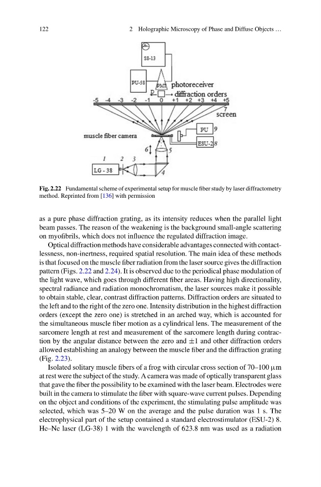
Fig. 2.22 Fundamental scheme of experimental setup for muscle fiber study by laser diffractometry
method. Reprinted from [136] with permission
as a pure phase diffraction grating, as its intensity reduces when the parallel light
beam passes. The reason of the weakening is the background small-angle scattering
on myofibrils, which does not influence the regulated diffraction image.
Optical diffraction methods have considerable advantages connected with contactlessness, non-inertness, required spatial resolution. The main idea of these methods
is that focused on the muscle fiber radiation from the laser source gives the diffraction
pattern (Figs. 2.22 and 2.24). It is observed due to the periodical phase modulation of
the light wave, which goes through different fiber areas. Having high directionality,
spectral radiance and radiation monochromatism, the laser sources make it possible
to obtain stable, clear, contrast diffraction patterns. Diffraction orders are situated to
the left and to the right of the zero one. Intensity distribution in the highest diffraction
orders (except the zero one) is stretched in an arched way, which is accounted for
the simultaneous muscle fiber motion as a cylindrical lens. The measurement of the
sarcomere length at rest and measurement of the sarcomere length during contraction by the angular distance between the zero and ±1 and other diffraction orders
allowed establishing an analogy between the muscle fiber and the diffraction grating
(Fig. 2.23).
Isolated solitary muscle fibers of a frog with circular cross section of 70–100 μm
at rest were the subject of the study. A camera was made of optically transparent glass
that gave the fiber the possibility to be examined with the laser beam. Electrodes were
built in the camera to stimulate the fiber with square-wave current pulses. Depending
on the object and conditions of the experiment, the stimulating pulse amplitude was
selected, which was 5–20 W on the average and the pulse duration was 1 s. The
electrophysical part of the setup contained a standard electrostimulator (ESU-2) 8.
He–Ne laser (LG-38) 1 with the wavelength of 623.8 nm was used as a radiation
2 Holographic Microscopy of Phase and Diffuse Objects …
Fig. 2.22 Fundamental scheme of experimental setup for muscle fiber study by laser diffractometry
method. Reprinted from [136] with permission
as a pure phase diffraction grating, as its intensity reduces when the parallel light
beam passes. The reason of the weakening is the background small-angle scattering
on myofibrils, which does not influence the regulated diffraction image.
Optical diffraction methods have considerable advantages connected with contactlessness, non-inertness, required spatial resolution. The main idea of these methods
is that focused on the muscle fiber radiation from the laser source gives the diffraction
pattern (Figs. 2.22 and 2.24). It is observed due to the periodical phase modulation of
the light wave, which goes through different fiber areas. Having high directionality,
spectral radiance and radiation monochromatism, the laser sources make it possible
to obtain stable, clear, contrast diffraction patterns. Diffraction orders are situated to
the left and to the right of the zero one. Intensity distribution in the highest diffraction
orders (except the zero one) is stretched in an arched way, which is accounted for
the simultaneous muscle fiber motion as a cylindrical lens. The measurement of the
sarcomere length at rest and measurement of the sarcomere length during contraction by the angular distance between the zero and ±1 and other diffraction orders
allowed establishing an analogy between the muscle fiber and the diffraction grating
(Fig. 2.23).
Isolated solitary muscle fibers of a frog with circular cross section of 70–100 μm
at rest were the subject of the study. A camera was made of optically transparent glass
that gave the fiber the possibility to be examined with the laser beam. Electrodes were
built in the camera to stimulate the fiber with square-wave current pulses. Depending
on the object and conditions of the experiment, the stimulating pulse amplitude was
selected, which was 5–20 W on the average and the pulse duration was 1 s. The
electrophysical part of the setup contained a standard electrostimulator (ESU-2) 8.
He–Ne laser (LG-38) 1 with the wavelength of 623.8 nm was used as a radiation