

98
2 Holographic Microscopy of Phase and Diffuse Objects …
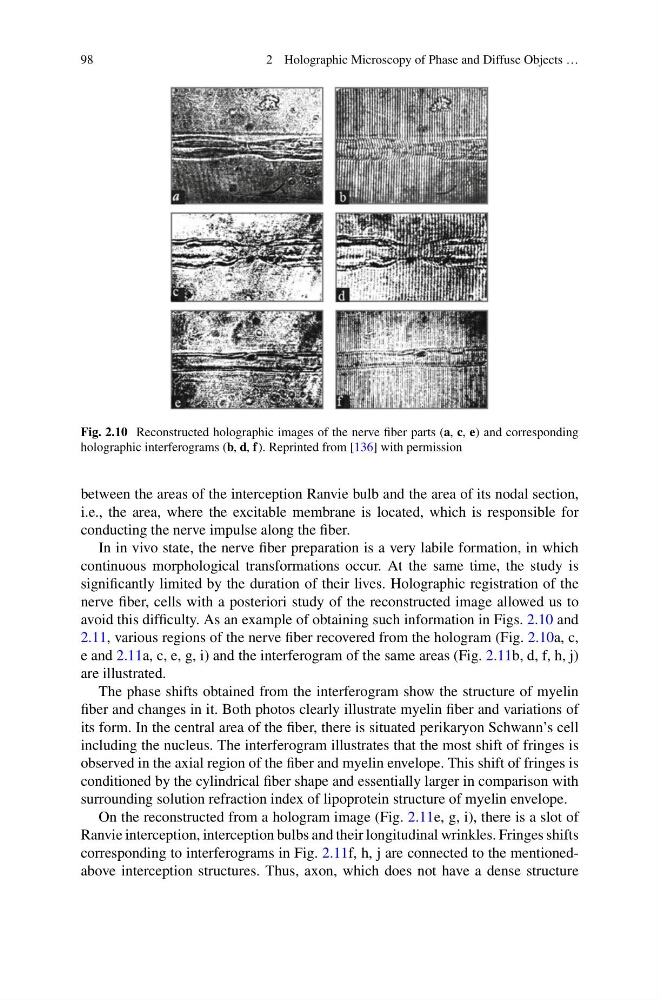
Fig. 2.10 Reconstructed holographic images of the nerve fiber parts (a, c, e) and corresponding
holographic interferograms (b, d, f). Reprinted from [136] with permission
between the areas of the interception Ranvie bulb and the area of its nodal section,
i.e., the area, where the excitable membrane is located, which is responsible for
conducting the nerve impulse along the fiber.
In in vivo state, the nerve fiber preparation is a very labile formation, in which
continuous morphological transformations occur. At the same time, the study is
significantly limited by the duration of their lives. Holographic registration of the
nerve fiber, cells with a posteriori study of the reconstructed image allowed us to
avoid this difficulty. As an example of obtaining such information in Figs. 2.10 and
2.11, various regions of the nerve fiber recovered from the hologram (Fig. 2.10a, c,
e and 2.11a, c, e, g, i) and the interferogram of the same areas (Fig. 2.11b, d, f, h, j)
are illustrated.
The phase shifts obtained from the interferogram show the structure of myelin
fiber and changes in it. Both photos clearly illustrate myelin fiber and variations of
its form. In the central area of the fiber, there is situated perikaryon Schwann’s cell
including the nucleus. The interferogram illustrates that the most shift of fringes is
observed in the axial region of the fiber and myelin envelope. This shift of fringes is
conditioned by the cylindrical fiber shape and essentially larger in comparison with
surrounding solution refraction index of lipoprotein structure of myelin envelope.
On the reconstructed from a hologram image (Fig. 2.11e, g, i), there is a slot of
Ranvie interception, interception bulbs and their longitudinal wrinkles. Fringes shifts
corresponding to interferograms in Fig. 2.11f, h, j are connected to the mentionedabove interception structures. Thus, axon, which does not have a dense structure
2 Holographic Microscopy of Phase and Diffuse Objects …
Fig. 2.10 Reconstructed holographic images of the nerve fiber parts (a, c, e) and corresponding
holographic interferograms (b, d, f). Reprinted from [136] with permission
between the areas of the interception Ranvie bulb and the area of its nodal section,
i.e., the area, where the excitable membrane is located, which is responsible for
conducting the nerve impulse along the fiber.
In in vivo state, the nerve fiber preparation is a very labile formation, in which
continuous morphological transformations occur. At the same time, the study is
significantly limited by the duration of their lives. Holographic registration of the
nerve fiber, cells with a posteriori study of the reconstructed image allowed us to
avoid this difficulty. As an example of obtaining such information in Figs. 2.10 and
2.11, various regions of the nerve fiber recovered from the hologram (Fig. 2.10a, c,
e and 2.11a, c, e, g, i) and the interferogram of the same areas (Fig. 2.11b, d, f, h, j)
are illustrated.
The phase shifts obtained from the interferogram show the structure of myelin
fiber and changes in it. Both photos clearly illustrate myelin fiber and variations of
its form. In the central area of the fiber, there is situated perikaryon Schwann’s cell
including the nucleus. The interferogram illustrates that the most shift of fringes is
observed in the axial region of the fiber and myelin envelope. This shift of fringes is
conditioned by the cylindrical fiber shape and essentially larger in comparison with
surrounding solution refraction index of lipoprotein structure of myelin envelope.
On the reconstructed from a hologram image (Fig. 2.11e, g, i), there is a slot of
Ranvie interception, interception bulbs and their longitudinal wrinkles. Fringes shifts
corresponding to interferograms in Fig. 2.11f, h, j are connected to the mentionedabove interception structures. Thus, axon, which does not have a dense structure