

2.2 Holographic Study of Structural and Functional Characteristics …
81
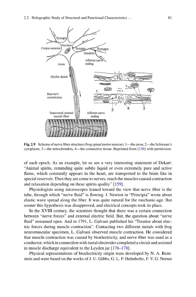
Fig. 2.9 Scheme of nerve fiber structure (frog spinal motor neuron): 1—the axon, 2—the Schwann’s
cytoplasm, 3—the mitochondria, 4—the connective tissue. Reprinted from [136] with permission
of each epoch. As an example, let us see a very interesting statement of Dekart:
“Animal spirits, reminding quite subtle liquid or even extremely pure and active
flame, which constantly appears in the heart, are transported to the brain like in
special reservoir. Then they are come to nerves, reach the muscles caused contraction
and relaxation depending on these spirits quality” [159].
Physiologists using microscopes leaned toward the view that nerve fiber is the
tube, through which “nerve fluid” is flowing. I. Newton in “Principia” wrote about
elastic wave spread along the fiber. It was quite natural for the mechanic-age. But
sooner this hypothesis was disapproved, and electrical concepts took its place.
In the XVIII century, the scientists thought that there was a certain connection
between “nerve forces” and external electric field. But, the question about “nerve
fluid” remained open. And in 1791, L. Galvani published his “Treatise about electric forces during muscle contraction”. Contacting two different metals with frog
neuromuscular specimen, L. Galvani observed muscle contraction. He considered
that muscle contraction was caused by bioelectricity, and nerve fiber was used as a
conductor, which in connection with metal electrodes completed a circuit and assisted
in muscle discharge equivalent to the Leyden jar [176–178].
Physical representations of bioelectricity origin were developed by N. A. Bernstein and were based on the works of J. U. Gibbs, G. L. F. Helmholtz, F. V. G. Nernst
81
Fig. 2.9 Scheme of nerve fiber structure (frog spinal motor neuron): 1—the axon, 2—the Schwann’s
cytoplasm, 3—the mitochondria, 4—the connective tissue. Reprinted from [136] with permission
of each epoch. As an example, let us see a very interesting statement of Dekart:
“Animal spirits, reminding quite subtle liquid or even extremely pure and active
flame, which constantly appears in the heart, are transported to the brain like in
special reservoir. Then they are come to nerves, reach the muscles caused contraction
and relaxation depending on these spirits quality” [159].
Physiologists using microscopes leaned toward the view that nerve fiber is the
tube, through which “nerve fluid” is flowing. I. Newton in “Principia” wrote about
elastic wave spread along the fiber. It was quite natural for the mechanic-age. But
sooner this hypothesis was disapproved, and electrical concepts took its place.
In the XVIII century, the scientists thought that there was a certain connection
between “nerve forces” and external electric field. But, the question about “nerve
fluid” remained open. And in 1791, L. Galvani published his “Treatise about electric forces during muscle contraction”. Contacting two different metals with frog
neuromuscular specimen, L. Galvani observed muscle contraction. He considered
that muscle contraction was caused by bioelectricity, and nerve fiber was used as a
conductor, which in connection with metal electrodes completed a circuit and assisted
in muscle discharge equivalent to the Leyden jar [176–178].
Physical representations of bioelectricity origin were developed by N. A. Bernstein and were based on the works of J. U. Gibbs, G. L. F. Helmholtz, F. V. G. Nernst