

10.3 The Eight-Layer Inversion Algorithm
255
Coil
Vessel Wall
1
σ
σ
σ
σ
σ
σ
σ
σ
7
8
2
3
4
5
6
Lipid Core : σ = 0.025
0
−0.85
−1.7
−2.0
L
(mm)
Blood : σ = 0.70
: σ = 0.58
Coil
Vessel Wall
1
σ
σ
σ
σ
σ
σ
σ
σ
7
8
2
3
4
5
6
Lipid Core : σ = 0.025
0
−1.7
−2.0
L
(mm)
−0.96
Blood : σ = 0.70
: σ = 0.58
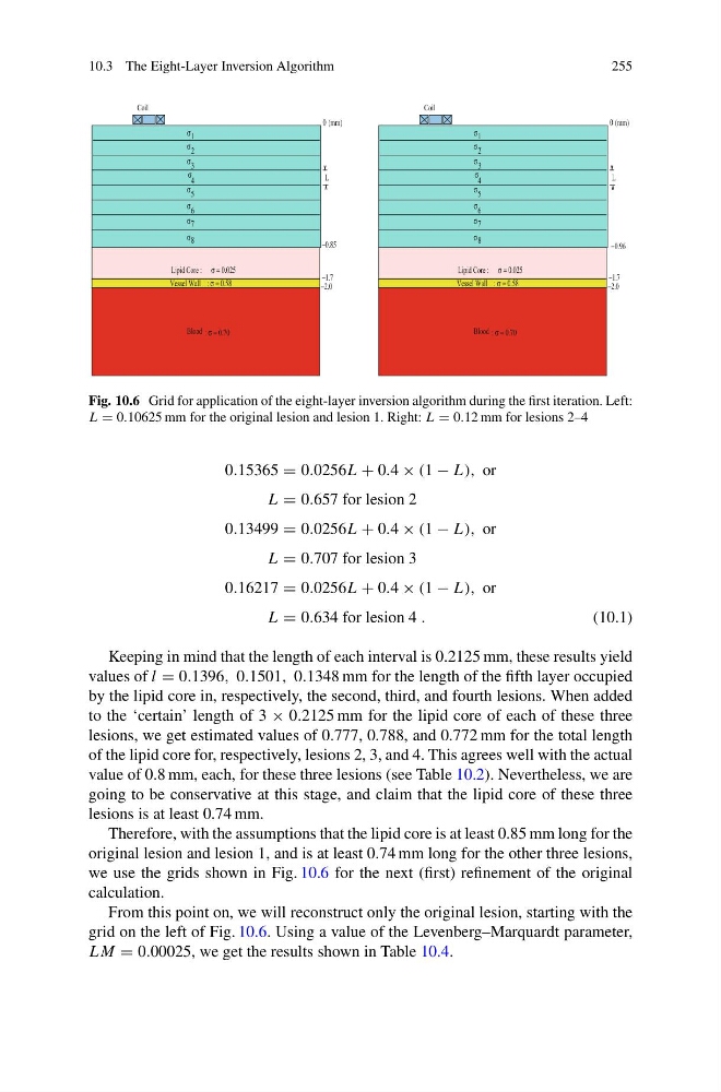
Fig. 10.6 Grid for application of the eight-layer inversion algorithm during the first iteration. Left:
L = 0.10625 mm for the original lesion and lesion 1. Right: L = 0.12 mm for lesions 2–4
0.15365 = 0.0256L + 0.4 × (1 − L), or
L = 0.657 for lesion 2
0.13499 = 0.0256L + 0.4 × (1 − L), or
L = 0.707 for lesion 3
0.16217 = 0.0256L + 0.4 × (1 − L), or
L = 0.634 for lesion 4 .
(10.1)
Keeping in mind that the length of each interval is 0.2125 mm, these results yield
values of l = 0.1396, 0.1501, 0.1348 mm for the length of the fifth layer occupied
by the lipid core in, respectively, the second, third, and fourth lesions. When added
to the ‘certain’ length of 3 × 0.2125 mm for the lipid core of each of these three
lesions, we get estimated values of 0.777, 0.788, and 0.772 mm for the total length
of the lipid core for, respectively, lesions 2, 3, and 4. This agrees well with the actual
value of 0.8 mm, each, for these three lesions (see Table 10.2). Nevertheless, we are
going to be conservative at this stage, and claim that the lipid core of these three
lesions is at least 0.74 mm.
Therefore, with the assumptions that the lipid core is at least 0.85 mm long for the
original lesion and lesion 1, and is at least 0.74 mm long for the other three lesions,
we use the grids shown in Fig. 10.6 for the next (first) refinement of the original
calculation.
From this point on, we will reconstruct only the original lesion, starting with the
grid on the left of Fig. 10.6. Using a value of the Levenberg–Marquardt parameter,
LM = 0.00025, we get the results shown in Table 10.4.
255
Coil
Vessel Wall
1
σ
σ
σ
σ
σ
σ
σ
σ
7
8
2
3
4
5
6
Lipid Core : σ = 0.025
0
−0.85
−1.7
−2.0
L
(mm)
Blood : σ = 0.70
: σ = 0.58
Coil
Vessel Wall
1
σ
σ
σ
σ
σ
σ
σ
σ
7
8
2
3
4
5
6
Lipid Core : σ = 0.025
0
−1.7
−2.0
L
(mm)
−0.96
Blood : σ = 0.70
: σ = 0.58
Fig. 10.6 Grid for application of the eight-layer inversion algorithm during the first iteration. Left:
L = 0.10625 mm for the original lesion and lesion 1. Right: L = 0.12 mm for lesions 2–4
0.15365 = 0.0256L + 0.4 × (1 − L), or
L = 0.657 for lesion 2
0.13499 = 0.0256L + 0.4 × (1 − L), or
L = 0.707 for lesion 3
0.16217 = 0.0256L + 0.4 × (1 − L), or
L = 0.634 for lesion 4 .
(10.1)
Keeping in mind that the length of each interval is 0.2125 mm, these results yield
values of l = 0.1396, 0.1501, 0.1348 mm for the length of the fifth layer occupied
by the lipid core in, respectively, the second, third, and fourth lesions. When added
to the ‘certain’ length of 3 × 0.2125 mm for the lipid core of each of these three
lesions, we get estimated values of 0.777, 0.788, and 0.772 mm for the total length
of the lipid core for, respectively, lesions 2, 3, and 4. This agrees well with the actual
value of 0.8 mm, each, for these three lesions (see Table 10.2). Nevertheless, we are
going to be conservative at this stage, and claim that the lipid core of these three
lesions is at least 0.74 mm.
Therefore, with the assumptions that the lipid core is at least 0.85 mm long for the
original lesion and lesion 1, and is at least 0.74 mm long for the other three lesions,
we use the grids shown in Fig. 10.6 for the next (first) refinement of the original
calculation.
From this point on, we will reconstruct only the original lesion, starting with the
grid on the left of Fig. 10.6. Using a value of the Levenberg–Marquardt parameter,
LM = 0.00025, we get the results shown in Table 10.4.