

256
10 Characterization of Atherosclerotic Lesions by Inversion of Eddy-. . .
Table 10.4 Results of
application of eight-layer
inversion algorithm using
Fig. 10.6. The
Levenberg–Marquardt
parameter is 0.00025
Lesion no.
Layer Upper boundary (mm) Original
1
0
0.079456
2
−0.10625
0.28272
3
−0.2125
0.34385
4
−0.31875
0.36084
5
−0.425
0.3445
6
−0.53125
0.29836
7
−0.6375
0.22236
8
−0.74375
0.11789
Coil
Vessel Wall
1
σ
σ
σ
σ
σ
σ
σ
σ
7
8
2
3
4
5
6
Lipid Core : σ = 0.025
−1.7
−2.0
L
0 (mm)
−0.69345
Blood : σ = 0.70
: σ = 0.58
Calcium : σ = 0.08
−0.10625
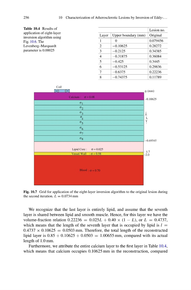
Fig. 10.7 Grid for application of the eight-layer inversion algorithm to the original lesion during
the second iteration. L = 0.0734 mm
We recognize that the last layer is entirely lipid, and assume that the seventh
layer is shared between lipid and smooth muscle. Hence, for this layer we have the
volume-fraction relation 0.22236 = 0.025L + 0.40 × (1 − L), or L = 0.4737,
which means that the length of the seventh layer that is occupied by lipid is l =
0.4737 × 0.10625 = 0.0503 mm. Therefore, the total length of the reconstructed
lipid layer is 0.85 + 0.10625 + 0.0503 = 1.00655 mm, compared with its actual
length of 1.0 mm.
Furthermore, we attribute the entire calcium layer to the first layer in Table 10.4,
which means that calcium occupies 0.10625 mm in the reconstruction, compared
10 Characterization of Atherosclerotic Lesions by Inversion of Eddy-. . .
Table 10.4 Results of
application of eight-layer
inversion algorithm using
Fig. 10.6. The
Levenberg–Marquardt
parameter is 0.00025
Lesion no.
Layer Upper boundary (mm) Original
1
0
0.079456
2
−0.10625
0.28272
3
−0.2125
0.34385
4
−0.31875
0.36084
5
−0.425
0.3445
6
−0.53125
0.29836
7
−0.6375
0.22236
8
−0.74375
0.11789
Coil
Vessel Wall
1
σ
σ
σ
σ
σ
σ
σ
σ
7
8
2
3
4
5
6
Lipid Core : σ = 0.025
−1.7
−2.0
L
0 (mm)
−0.69345
Blood : σ = 0.70
: σ = 0.58
Calcium : σ = 0.08
−0.10625
Fig. 10.7 Grid for application of the eight-layer inversion algorithm to the original lesion during
the second iteration. L = 0.0734 mm
We recognize that the last layer is entirely lipid, and assume that the seventh
layer is shared between lipid and smooth muscle. Hence, for this layer we have the
volume-fraction relation 0.22236 = 0.025L + 0.40 × (1 − L), or L = 0.4737,
which means that the length of the seventh layer that is occupied by lipid is l =
0.4737 × 0.10625 = 0.0503 mm. Therefore, the total length of the reconstructed
lipid layer is 0.85 + 0.10625 + 0.0503 = 1.00655 mm, compared with its actual
length of 1.0 mm.
Furthermore, we attribute the entire calcium layer to the first layer in Table 10.4,
which means that calcium occupies 0.10625 mm in the reconstruction, compared