

4.6 Swarms and Colonies
85
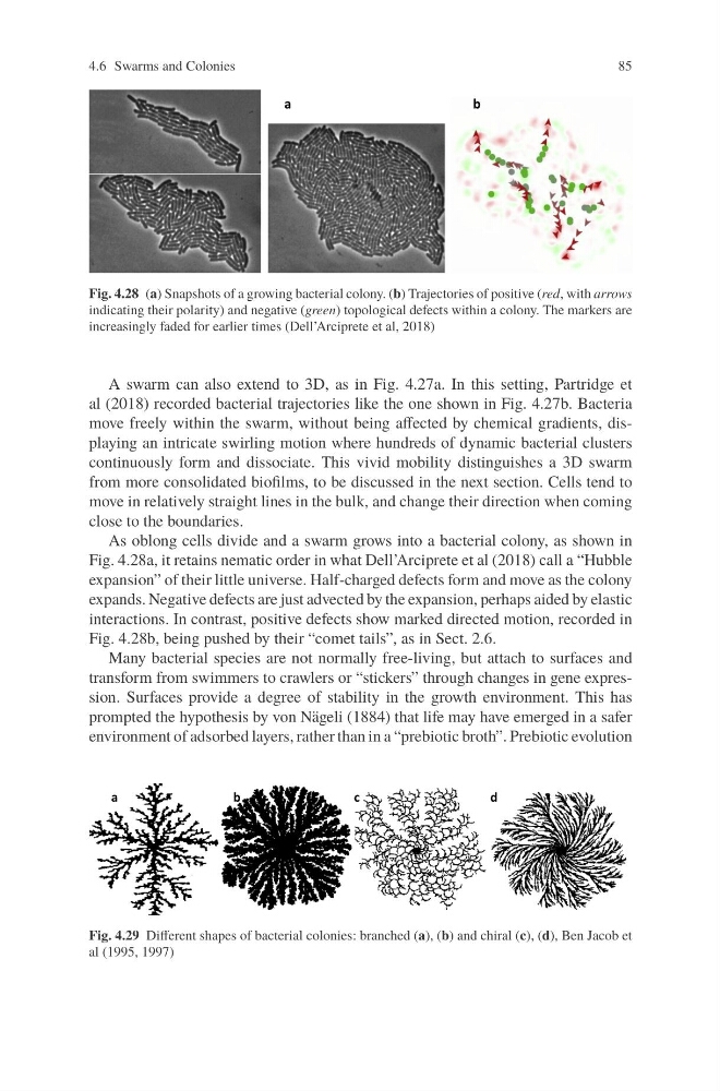
Fig. 4.28 (a) Snapshots of a growing bacterial colony. (b) Trajectories of positive (red, with arrows
indicating their polarity) and negative (green) topological defects within a colony. The markers are
increasingly faded for earlier times (Dell’Arciprete et al, 2018)
A swarm can also extend to 3D, as in Fig. 4.27a. In this setting, Partridge et
al (2018) recorded bacterial trajectories like the one shown in Fig. 4.27b. Bacteria
move freely within the swarm, without being affected by chemical gradients, displaying an intricate swirling motion where hundreds of dynamic bacterial clusters
continuously form and dissociate. This vivid mobility distinguishes a 3D swarm
from more consolidated biofilms, to be discussed in the next section. Cells tend to
move in relatively straight lines in the bulk, and change their direction when coming
close to the boundaries.
As oblong cells divide and a swarm grows into a bacterial colony, as shown in
Fig. 4.28a, it retains nematic order in what Dell’Arciprete et al (2018) call a “Hubble
expansion” of their little universe. Half-charged defects form and move as the colony
expands. Negative defects are just advected by the expansion, perhaps aided by elastic
interactions. In contrast, positive defects show marked directed motion, recorded in
Fig. 4.28b, being pushed by their “comet tails”, as in Sect. 2.6.
Many bacterial species are not normally free-living, but attach to surfaces and
transform from swimmers to crawlers or “stickers” through changes in gene expression. Surfaces provide a degree of stability in the growth environment. This has
prompted the hypothesis by von Nägeli (1884) that life may have emerged in a safer
environment of adsorbed layers, rather than in a “prebiotic broth”. Prebiotic evolution
Fig. 4.29 Different shapes of bacterial colonies: branched (a), (b) and chiral (c), (d), Ben Jacob et
al (1995, 1997)
85
Fig. 4.28 (a) Snapshots of a growing bacterial colony. (b) Trajectories of positive (red, with arrows
indicating their polarity) and negative (green) topological defects within a colony. The markers are
increasingly faded for earlier times (Dell’Arciprete et al, 2018)
A swarm can also extend to 3D, as in Fig. 4.27a. In this setting, Partridge et
al (2018) recorded bacterial trajectories like the one shown in Fig. 4.27b. Bacteria
move freely within the swarm, without being affected by chemical gradients, displaying an intricate swirling motion where hundreds of dynamic bacterial clusters
continuously form and dissociate. This vivid mobility distinguishes a 3D swarm
from more consolidated biofilms, to be discussed in the next section. Cells tend to
move in relatively straight lines in the bulk, and change their direction when coming
close to the boundaries.
As oblong cells divide and a swarm grows into a bacterial colony, as shown in
Fig. 4.28a, it retains nematic order in what Dell’Arciprete et al (2018) call a “Hubble
expansion” of their little universe. Half-charged defects form and move as the colony
expands. Negative defects are just advected by the expansion, perhaps aided by elastic
interactions. In contrast, positive defects show marked directed motion, recorded in
Fig. 4.28b, being pushed by their “comet tails”, as in Sect. 2.6.
Many bacterial species are not normally free-living, but attach to surfaces and
transform from swimmers to crawlers or “stickers” through changes in gene expression. Surfaces provide a degree of stability in the growth environment. This has
prompted the hypothesis by von Nägeli (1884) that life may have emerged in a safer
environment of adsorbed layers, rather than in a “prebiotic broth”. Prebiotic evolution
Fig. 4.29 Different shapes of bacterial colonies: branched (a), (b) and chiral (c), (d), Ben Jacob et
al (1995, 1997)