

86
4 Motion of Microorganisms
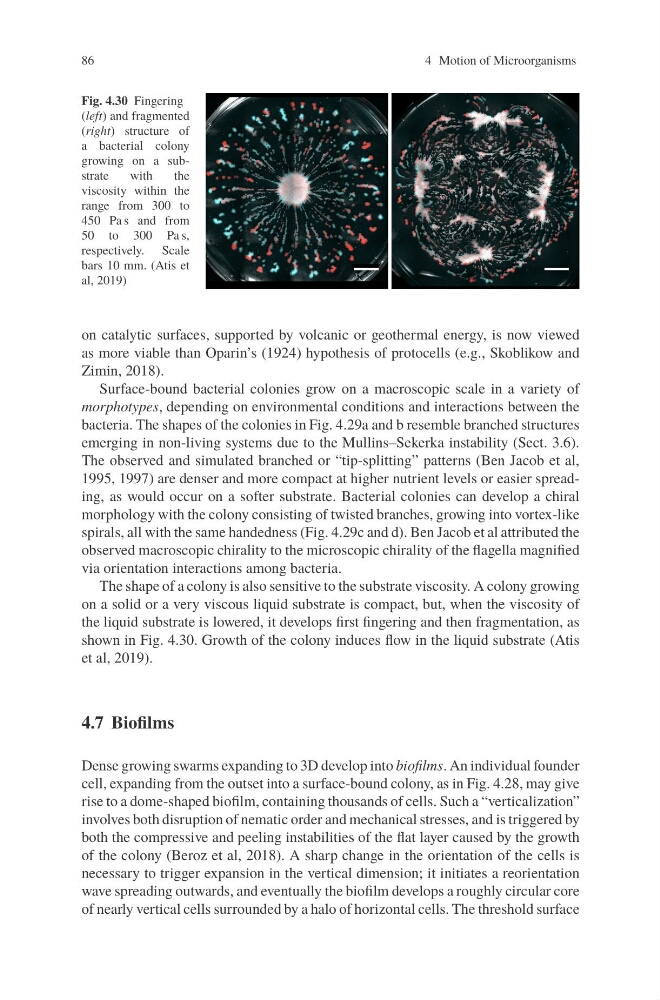
Fig. 4.30 Fingering
(left) and fragmented
(right) structure of
a bacterial colony
growing on a substrate
with
the
viscosity within the
range from 300 to
450 Pa s and from
50 to 300 Pa s,
respectively. Scale
bars 10 mm. (Atis et
al, 2019)
on catalytic surfaces, supported by volcanic or geothermal energy, is now viewed
as more viable than Oparin’s (1924) hypothesis of protocells (e.g., Skoblikow and
Zimin, 2018).
Surface-bound bacterial colonies grow on a macroscopic scale in a variety of
morphotypes, depending on environmental conditions and interactions between the
bacteria. The shapes of the colonies in Fig. 4.29a and b resemble branched structures
emerging in non-living systems due to the Mullins–Sekerka instability (Sect. 3.6).
The observed and simulated branched or “tip-splitting” patterns (Ben Jacob et al,
1995, 1997) are denser and more compact at higher nutrient levels or easier spreading, as would occur on a softer substrate. Bacterial colonies can develop a chiral
morphology with the colony consisting of twisted branches, growing into vortex-like
spirals, all with the same handedness (Fig. 4.29c and d). Ben Jacob et al attributed the
observed macroscopic chirality to the microscopic chirality of the flagella magnified
via orientation interactions among bacteria.
The shape of a colony is also sensitive to the substrate viscosity. A colony growing
on a solid or a very viscous liquid substrate is compact, but, when the viscosity of
the liquid substrate is lowered, it develops first fingering and then fragmentation, as
shown in Fig. 4.30. Growth of the colony induces flow in the liquid substrate (Atis
et al, 2019).
4.7 Biofilms
Dense growing swarms expanding to 3D develop into biofilms. An individual founder
cell, expanding from the outset into a surface-bound colony, as in Fig. 4.28, may give
rise to a dome-shaped biofilm, containing thousands of cells. Such a “verticalization”
involves both disruption of nematic order and mechanical stresses, and is triggered by
both the compressive and peeling instabilities of the flat layer caused by the growth
of the colony (Beroz et al, 2018). A sharp change in the orientation of the cells is
necessary to trigger expansion in the vertical dimension; it initiates a reorientation
wave spreading outwards, and eventually the biofilm develops a roughly circular core
of nearly vertical cells surrounded by a halo of horizontal cells. The threshold surface
4 Motion of Microorganisms
Fig. 4.30 Fingering
(left) and fragmented
(right) structure of
a bacterial colony
growing on a substrate
with
the
viscosity within the
range from 300 to
450 Pa s and from
50 to 300 Pa s,
respectively. Scale
bars 10 mm. (Atis et
al, 2019)
on catalytic surfaces, supported by volcanic or geothermal energy, is now viewed
as more viable than Oparin’s (1924) hypothesis of protocells (e.g., Skoblikow and
Zimin, 2018).
Surface-bound bacterial colonies grow on a macroscopic scale in a variety of
morphotypes, depending on environmental conditions and interactions between the
bacteria. The shapes of the colonies in Fig. 4.29a and b resemble branched structures
emerging in non-living systems due to the Mullins–Sekerka instability (Sect. 3.6).
The observed and simulated branched or “tip-splitting” patterns (Ben Jacob et al,
1995, 1997) are denser and more compact at higher nutrient levels or easier spreading, as would occur on a softer substrate. Bacterial colonies can develop a chiral
morphology with the colony consisting of twisted branches, growing into vortex-like
spirals, all with the same handedness (Fig. 4.29c and d). Ben Jacob et al attributed the
observed macroscopic chirality to the microscopic chirality of the flagella magnified
via orientation interactions among bacteria.
The shape of a colony is also sensitive to the substrate viscosity. A colony growing
on a solid or a very viscous liquid substrate is compact, but, when the viscosity of
the liquid substrate is lowered, it develops first fingering and then fragmentation, as
shown in Fig. 4.30. Growth of the colony induces flow in the liquid substrate (Atis
et al, 2019).
4.7 Biofilms
Dense growing swarms expanding to 3D develop into biofilms. An individual founder
cell, expanding from the outset into a surface-bound colony, as in Fig. 4.28, may give
rise to a dome-shaped biofilm, containing thousands of cells. Such a “verticalization”
involves both disruption of nematic order and mechanical stresses, and is triggered by
both the compressive and peeling instabilities of the flat layer caused by the growth
of the colony (Beroz et al, 2018). A sharp change in the orientation of the cells is
necessary to trigger expansion in the vertical dimension; it initiates a reorientation
wave spreading outwards, and eventually the biofilm develops a roughly circular core
of nearly vertical cells surrounded by a halo of horizontal cells. The threshold surface