

78
4 Motion of Microorganisms
bacteria belonging to this strain are marked green, shows their rather rapid (on the
scale of half an hour) aggregation from a dilute suspension.
Crowding in dense suspensions inhibits chemotactic sensing, leaving bacteria less
space for runs that would allow them to feel the gradient, although the swimming
speed may increase slightly due to collective entrainment. As the cell is swimming,
it monitors the change in chemoattractant concentration within a few seconds, to
decide whether to tumble. If during this time the direction in which the cell swims
has changed significantly due to steric interactions with close neighbors, the decision
becomes less relevant, thereby making the bacterial chemotaxis strategy inefficient
(Colin et al, 2019).
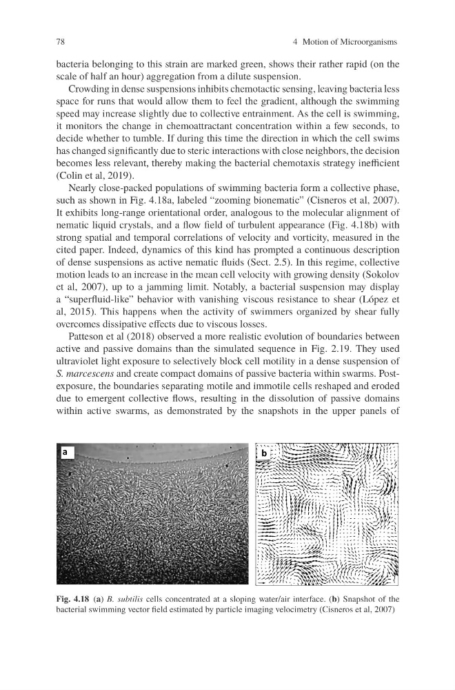
Nearly close-packed populations of swimming bacteria form a collective phase,
such as shown in Fig. 4.18a, labeled “zooming bionematic” (Cisneros et al, 2007).
It exhibits long-range orientational order, analogous to the molecular alignment of
nematic liquid crystals, and a flow field of turbulent appearance (Fig. 4.18b) with
strong spatial and temporal correlations of velocity and vorticity, measured in the
cited paper. Indeed, dynamics of this kind has prompted a continuous description
of dense suspensions as active nematic fluids (Sect. 2.5). In this regime, collective
motion leads to an increase in the mean cell velocity with growing density (Sokolov
et al, 2007), up to a jamming limit. Notably, a bacterial suspension may display
a “superfluid-like” behavior with vanishing viscous resistance to shear (López et
al, 2015). This happens when the activity of swimmers organized by shear fully
overcomes dissipative effects due to viscous losses.
Patteson et al (2018) observed a more realistic evolution of boundaries between
active and passive domains than the simulated sequence in Fig. 2.19. They used
ultraviolet light exposure to selectively block cell motility in a dense suspension of
S. marcescens and create compact domains of passive bacteria within swarms. Postexposure, the boundaries separating motile and immotile cells reshaped and eroded
due to emergent collective flows, resulting in the dissolution of passive domains
within active swarms, as demonstrated by the snapshots in the upper panels of
a
a
a
b
Fig. 4.18 (a) B. subtilis cells concentrated at a sloping water/air interface. (b) Snapshot of the
bacterial swimming vector field estimated by particle imaging velocimetry (Cisneros et al, 2007)
4 Motion of Microorganisms
bacteria belonging to this strain are marked green, shows their rather rapid (on the
scale of half an hour) aggregation from a dilute suspension.
Crowding in dense suspensions inhibits chemotactic sensing, leaving bacteria less
space for runs that would allow them to feel the gradient, although the swimming
speed may increase slightly due to collective entrainment. As the cell is swimming,
it monitors the change in chemoattractant concentration within a few seconds, to
decide whether to tumble. If during this time the direction in which the cell swims
has changed significantly due to steric interactions with close neighbors, the decision
becomes less relevant, thereby making the bacterial chemotaxis strategy inefficient
(Colin et al, 2019).
Nearly close-packed populations of swimming bacteria form a collective phase,
such as shown in Fig. 4.18a, labeled “zooming bionematic” (Cisneros et al, 2007).
It exhibits long-range orientational order, analogous to the molecular alignment of
nematic liquid crystals, and a flow field of turbulent appearance (Fig. 4.18b) with
strong spatial and temporal correlations of velocity and vorticity, measured in the
cited paper. Indeed, dynamics of this kind has prompted a continuous description
of dense suspensions as active nematic fluids (Sect. 2.5). In this regime, collective
motion leads to an increase in the mean cell velocity with growing density (Sokolov
et al, 2007), up to a jamming limit. Notably, a bacterial suspension may display
a “superfluid-like” behavior with vanishing viscous resistance to shear (López et
al, 2015). This happens when the activity of swimmers organized by shear fully
overcomes dissipative effects due to viscous losses.
Patteson et al (2018) observed a more realistic evolution of boundaries between
active and passive domains than the simulated sequence in Fig. 2.19. They used
ultraviolet light exposure to selectively block cell motility in a dense suspension of
S. marcescens and create compact domains of passive bacteria within swarms. Postexposure, the boundaries separating motile and immotile cells reshaped and eroded
due to emergent collective flows, resulting in the dissolution of passive domains
within active swarms, as demonstrated by the snapshots in the upper panels of
a
a
a
b
Fig. 4.18 (a) B. subtilis cells concentrated at a sloping water/air interface. (b) Snapshot of the
bacterial swimming vector field estimated by particle imaging velocimetry (Cisneros et al, 2007)