

3.6 Imitated Cells
59
A requisite element of Oparin’s hypothesis is a membrane keeping the content
of protocells intact. Such a barrier separating replicating biomolecules from the
environment is essential for specialization and competition between cells. It could
have emerged quite naturally at the dawn of life, built of surfactant molecules present
in a chaotic primordial soup of organic matter. Ruiz-Herrero et al (2019), instead of
focusing on a basic chemical metabolism, built up a model that couples membrane
growth and fluid permeation. The surface of a vesicle may rapidly increase by
addition of surfactant molecules, but its volume could grow slowly, as it is limited by
the permeability of the membrane and may be counteracted osmotically, depending
on the balance of solute concentrations inside and outside the micelle. In this model,
further evolution depends on two factors: the rate of mechanical relaxation and the
preferred spontaneous curvature. It unfolds in a similar way to the evolution in the
model of Zwicker et al when the mechanical relaxation is fast and the preferred
curvature is positive: as the surface to volume ratio increases, the vesicle elongates,
becomes mechanically unstable, and breaks into smaller vesicles (Fig. 3.22a). There
is also a technical problem here related to the division event. This behavior may
be irrelevant for the question of the origin of life (where Oparin’s hypothesis is no
longer viewed as viable) but it is consistent with observations of cell deformation
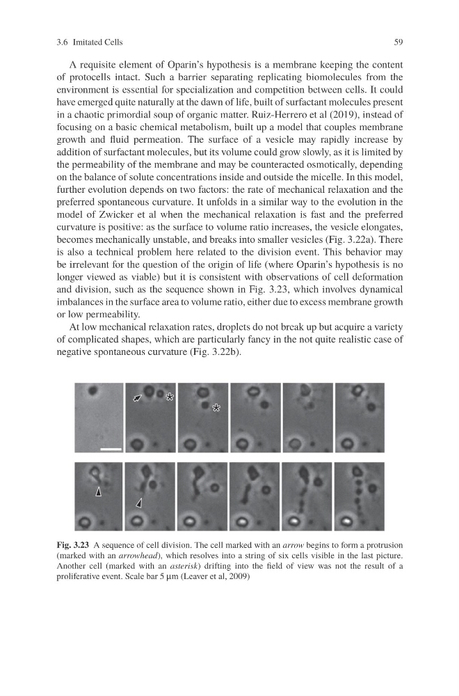
and division, such as the sequence shown in Fig. 3.23, which involves dynamical
imbalances in the surface area to volume ratio, either due to excess membrane growth
or low permeability.
At low mechanical relaxation rates, droplets do not break up but acquire a variety
of complicated shapes, which are particularly fancy in the not quite realistic case of
negative spontaneous curvature (Fig. 3.22b).
Fig. 3.23 A sequence of cell division. The cell marked with an arrow begins to form a protrusion
(marked with an arrowhead), which resolves into a string of six cells visible in the last picture.
Another cell (marked with an asterisk) drifting into the field of view was not the result of a
proliferative event. Scale bar 5 μm (Leaver et al, 2009)
59
A requisite element of Oparin’s hypothesis is a membrane keeping the content
of protocells intact. Such a barrier separating replicating biomolecules from the
environment is essential for specialization and competition between cells. It could
have emerged quite naturally at the dawn of life, built of surfactant molecules present
in a chaotic primordial soup of organic matter. Ruiz-Herrero et al (2019), instead of
focusing on a basic chemical metabolism, built up a model that couples membrane
growth and fluid permeation. The surface of a vesicle may rapidly increase by
addition of surfactant molecules, but its volume could grow slowly, as it is limited by
the permeability of the membrane and may be counteracted osmotically, depending
on the balance of solute concentrations inside and outside the micelle. In this model,
further evolution depends on two factors: the rate of mechanical relaxation and the
preferred spontaneous curvature. It unfolds in a similar way to the evolution in the
model of Zwicker et al when the mechanical relaxation is fast and the preferred
curvature is positive: as the surface to volume ratio increases, the vesicle elongates,
becomes mechanically unstable, and breaks into smaller vesicles (Fig. 3.22a). There
is also a technical problem here related to the division event. This behavior may
be irrelevant for the question of the origin of life (where Oparin’s hypothesis is no
longer viewed as viable) but it is consistent with observations of cell deformation
and division, such as the sequence shown in Fig. 3.23, which involves dynamical
imbalances in the surface area to volume ratio, either due to excess membrane growth
or low permeability.
At low mechanical relaxation rates, droplets do not break up but acquire a variety
of complicated shapes, which are particularly fancy in the not quite realistic case of
negative spontaneous curvature (Fig. 3.22b).
Fig. 3.23 A sequence of cell division. The cell marked with an arrow begins to form a protrusion
(marked with an arrowhead), which resolves into a string of six cells visible in the last picture.
Another cell (marked with an asterisk) drifting into the field of view was not the result of a
proliferative event. Scale bar 5 μm (Leaver et al, 2009)