

8.6 Remodeling
191
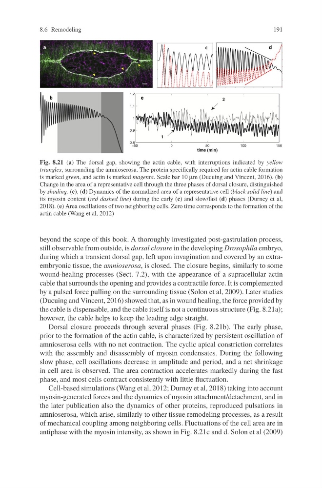
Fig. 8.21 (a) The dorsal gap, showing the actin cable, with interruptions indicated by yellow
triangles, surrounding the amnioserosa. The protein specifically required for actin cable formation
is marked green, and actin is marked magenta. Scale bar 10 μm (Ducuing and Vincent, 2016). (b)
Change in the area of a representative cell through the three phases of dorsal closure, distinguished
by shading. (c), (d) Dynamics of the normalized area of a representative cell (black solid line) and
its myosin content (red dashed line) during the early (c) and slow/fast (d) phases (Durney et al,
2018). (e) Area oscillations of two neighboring cells. Zero time corresponds to the formation of the
actin cable (Wang et al, 2012)
beyond the scope of this book. A thoroughly investigated post-gastrulation process,
still observable from outside, is dorsal closure in the developing Drosophila embryo,
during which a transient dorsal gap, left upon invagination and covered by an extraembryonic tissue, the amnioserosa, is closed. The closure begins, similarly to some
wound-healing processes (Sect. 7.2), with the appearance of a supracellular actin
cable that surrounds the opening and provides a contractile force. It is complemented
by a pulsed force pulling on the surrounding tissue (Solon et al, 2009). Later studies
(Ducuing and Vincent, 2016) showed that, as in wound healing, the force provided by
the cable is dispensable, and the cable itself is not a continuous structure (Fig. 8.21a);
however, the cable helps to keep the leading edge straight.
Dorsal closure proceeds through several phases (Fig. 8.21b). The early phase,
prior to the formation of the actin cable, is characterized by persistent oscillation of
amnioserosa cells with no net contraction. The cyclic apical constriction correlates
with the assembly and disassembly of myosin condensates. During the following
slow phase, cell oscillations decrease in amplitude and period, and a net shrinkage
in cell area is observed. The area contraction accelerates markedly during the fast
phase, and most cells contract consistently with little fluctuation.
Cell-based simulations (Wang et al, 2012; Durney et al, 2018) taking into account
myosin-generated forces and the dynamics of myosin attachment/detachment, and in
the later publication also the dynamics of other proteins, reproduced pulsations in
amnioserosa, which arise, similarly to other tissue remodeling processes, as a result
of mechanical coupling among neighboring cells. Fluctuations of the cell area are in
antiphase with the myosin intensity, as shown in Fig. 8.21c and d. Solon et al (2009)
191
Fig. 8.21 (a) The dorsal gap, showing the actin cable, with interruptions indicated by yellow
triangles, surrounding the amnioserosa. The protein specifically required for actin cable formation
is marked green, and actin is marked magenta. Scale bar 10 μm (Ducuing and Vincent, 2016). (b)
Change in the area of a representative cell through the three phases of dorsal closure, distinguished
by shading. (c), (d) Dynamics of the normalized area of a representative cell (black solid line) and
its myosin content (red dashed line) during the early (c) and slow/fast (d) phases (Durney et al,
2018). (e) Area oscillations of two neighboring cells. Zero time corresponds to the formation of the
actin cable (Wang et al, 2012)
beyond the scope of this book. A thoroughly investigated post-gastrulation process,
still observable from outside, is dorsal closure in the developing Drosophila embryo,
during which a transient dorsal gap, left upon invagination and covered by an extraembryonic tissue, the amnioserosa, is closed. The closure begins, similarly to some
wound-healing processes (Sect. 7.2), with the appearance of a supracellular actin
cable that surrounds the opening and provides a contractile force. It is complemented
by a pulsed force pulling on the surrounding tissue (Solon et al, 2009). Later studies
(Ducuing and Vincent, 2016) showed that, as in wound healing, the force provided by
the cable is dispensable, and the cable itself is not a continuous structure (Fig. 8.21a);
however, the cable helps to keep the leading edge straight.
Dorsal closure proceeds through several phases (Fig. 8.21b). The early phase,
prior to the formation of the actin cable, is characterized by persistent oscillation of
amnioserosa cells with no net contraction. The cyclic apical constriction correlates
with the assembly and disassembly of myosin condensates. During the following
slow phase, cell oscillations decrease in amplitude and period, and a net shrinkage
in cell area is observed. The area contraction accelerates markedly during the fast
phase, and most cells contract consistently with little fluctuation.
Cell-based simulations (Wang et al, 2012; Durney et al, 2018) taking into account
myosin-generated forces and the dynamics of myosin attachment/detachment, and in
the later publication also the dynamics of other proteins, reproduced pulsations in
amnioserosa, which arise, similarly to other tissue remodeling processes, as a result
of mechanical coupling among neighboring cells. Fluctuations of the cell area are in
antiphase with the myosin intensity, as shown in Fig. 8.21c and d. Solon et al (2009)