

8.5 Mechanotransduction
185
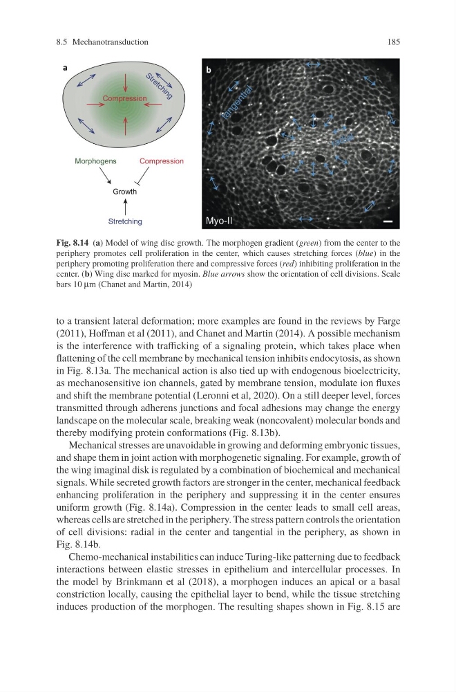
Fig. 8.14 (a) Model of wing disc growth. The morphogen gradient (green) from the center to the
periphery promotes cell proliferation in the center, which causes stretching forces (blue) in the
periphery promoting proliferation there and compressive forces (red) inhibiting proliferation in the
center. (b) Wing disc marked for myosin. Blue arrows show the orientation of cell divisions. Scale
bars 10 μm (Chanet and Martin, 2014)
to a transient lateral deformation; more examples are found in the reviews by Farge
(2011), Hoffman et al (2011), and Chanet and Martin (2014). A possible mechanism
is the interference with trafficking of a signaling protein, which takes place when
flattening of the cell membrane by mechanical tension inhibits endocytosis, as shown
in Fig. 8.13a. The mechanical action is also tied up with endogenous bioelectricity,
as mechanosensitive ion channels, gated by membrane tension, modulate ion fluxes
and shift the membrane potential (Leronni et al, 2020). On a still deeper level, forces
transmitted through adherens junctions and focal adhesions may change the energy
landscape on the molecular scale, breaking weak (noncovalent) molecular bonds and
thereby modifying protein conformations (Fig. 8.13b).
Mechanical stresses are unavoidable in growing and deforming embryonic tissues,
and shape them in joint action with morphogenetic signaling. For example, growth of
the wing imaginal disk is regulated by a combination of biochemical and mechanical
signals. While secreted growth factors are stronger in the center, mechanical feedback
enhancing proliferation in the periphery and suppressing it in the center ensures
uniform growth (Fig. 8.14a). Compression in the center leads to small cell areas,
whereas cells are stretched in the periphery. The stress pattern controls the orientation
of cell divisions: radial in the center and tangential in the periphery, as shown in
Fig. 8.14b.
Chemo-mechanical instabilities can induce Turing-like patterning due to feedback
interactions between elastic stresses in epithelium and intercellular processes. In
the model by Brinkmann et al (2018), a morphogen induces an apical or a basal
constriction locally, causing the epithelial layer to bend, while the tissue stretching
induces production of the morphogen. The resulting shapes shown in Fig. 8.15 are
185
Fig. 8.14 (a) Model of wing disc growth. The morphogen gradient (green) from the center to the
periphery promotes cell proliferation in the center, which causes stretching forces (blue) in the
periphery promoting proliferation there and compressive forces (red) inhibiting proliferation in the
center. (b) Wing disc marked for myosin. Blue arrows show the orientation of cell divisions. Scale
bars 10 μm (Chanet and Martin, 2014)
to a transient lateral deformation; more examples are found in the reviews by Farge
(2011), Hoffman et al (2011), and Chanet and Martin (2014). A possible mechanism
is the interference with trafficking of a signaling protein, which takes place when
flattening of the cell membrane by mechanical tension inhibits endocytosis, as shown
in Fig. 8.13a. The mechanical action is also tied up with endogenous bioelectricity,
as mechanosensitive ion channels, gated by membrane tension, modulate ion fluxes
and shift the membrane potential (Leronni et al, 2020). On a still deeper level, forces
transmitted through adherens junctions and focal adhesions may change the energy
landscape on the molecular scale, breaking weak (noncovalent) molecular bonds and
thereby modifying protein conformations (Fig. 8.13b).
Mechanical stresses are unavoidable in growing and deforming embryonic tissues,
and shape them in joint action with morphogenetic signaling. For example, growth of
the wing imaginal disk is regulated by a combination of biochemical and mechanical
signals. While secreted growth factors are stronger in the center, mechanical feedback
enhancing proliferation in the periphery and suppressing it in the center ensures
uniform growth (Fig. 8.14a). Compression in the center leads to small cell areas,
whereas cells are stretched in the periphery. The stress pattern controls the orientation
of cell divisions: radial in the center and tangential in the periphery, as shown in
Fig. 8.14b.
Chemo-mechanical instabilities can induce Turing-like patterning due to feedback
interactions between elastic stresses in epithelium and intercellular processes. In
the model by Brinkmann et al (2018), a morphogen induces an apical or a basal
constriction locally, causing the epithelial layer to bend, while the tissue stretching
induces production of the morphogen. The resulting shapes shown in Fig. 8.15 are