

166
7 Live Tissues
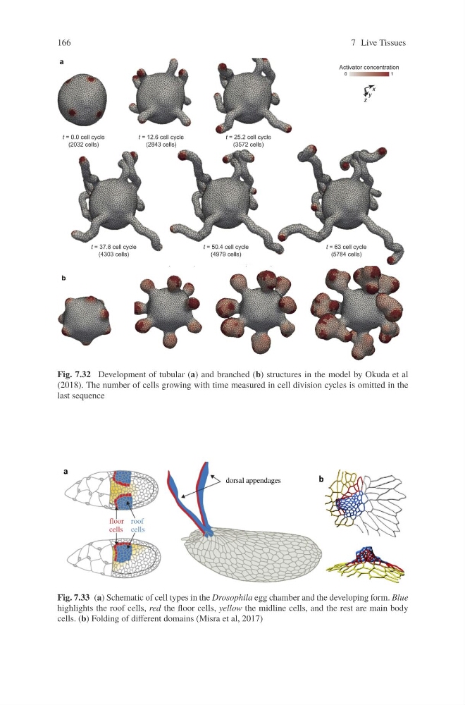
Fig. 7.32 Development of tubular (a) and branched (b) structures in the model by Okuda et al
(2018). The number of cells growing with time measured in cell division cycles is omitted in the
last sequence
Fig. 7.33 (a) Schematic of cell types in the Drosophila egg chamber and the developing form. Blue
highlights the roof cells, red the floor cells, yellow the midline cells, and the rest are main body
cells. (b) Folding of different domains (Misra et al, 2017)
7 Live Tissues
Fig. 7.32 Development of tubular (a) and branched (b) structures in the model by Okuda et al
(2018). The number of cells growing with time measured in cell division cycles is omitted in the
last sequence
Fig. 7.33 (a) Schematic of cell types in the Drosophila egg chamber and the developing form. Blue
highlights the roof cells, red the floor cells, yellow the midline cells, and the rest are main body
cells. (b) Folding of different domains (Misra et al, 2017)