

164
7 Live Tissues
enhanced actomyosin activity, as sketched in Fig. 7.29a. The resulting bending of
the epithelial layer is schematized in Fig. 7.29b.
In a pioneering study of chemo-mechanical interactions, Odell et al (1981) postulated, without specifying a molecular mechanism, that a sufficiently large extension
of the cellular cortex triggers restoring forces which are so strong that they cause it to
contract beyond their equilibrium configuration. The coordination of epithelial cell
shape changes is accompanied by the propagation of mechanical contraction waves.
Beloussov and Mittenthal (1992) generalized this kind of response as hyperrestoration, which they put forward as a fundamental property of a developing and growing
organism (although, of course, it is not expected to operate in all circumstances).
This response is apt to generate a wave of cell shape change propagating along an
epithelial layer. On this principle, Odell et al built up a 1D cell-based model of
epithelial invagination, one of the most important morphogenetic processes (more
on this in Sect. 8.6).
More complex 3D forms may be formed by apical constriction of a group of cells,
as in Fig. 7.29c. Another possible cause of bending is modulation of apical–basal
polarity (Fig. 7.29d and e) by shifting positions of adherens junctions (Wang et al,
2013).
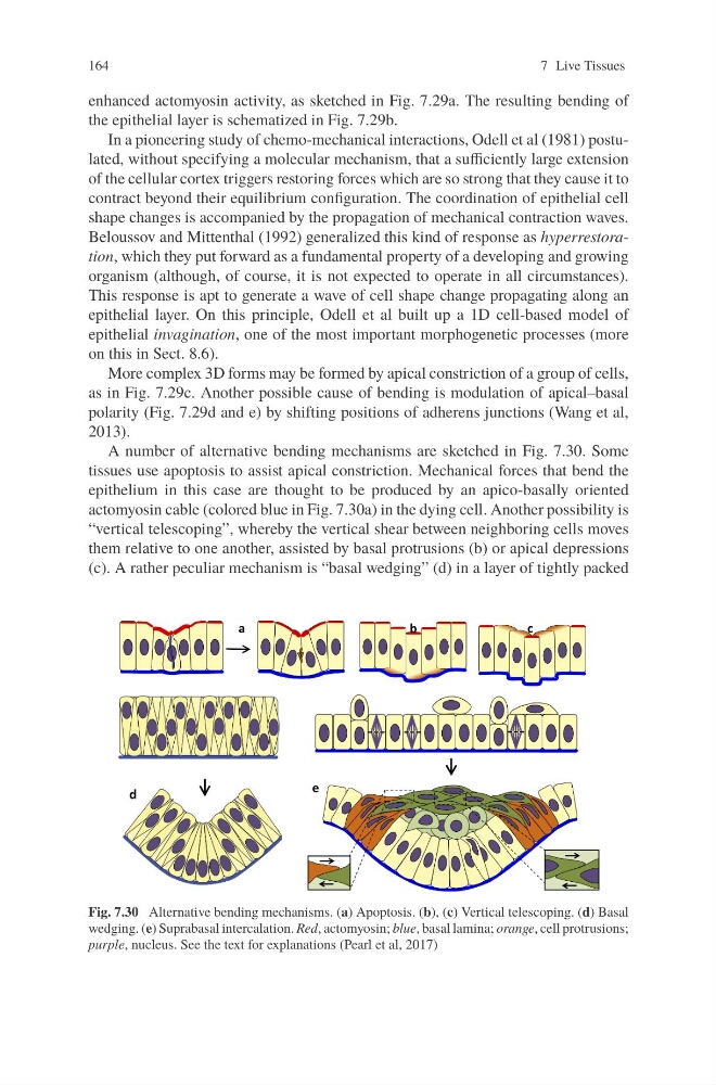
A number of alternative bending mechanisms are sketched in Fig. 7.30. Some
tissues use apoptosis to assist apical constriction. Mechanical forces that bend the
epithelium in this case are thought to be produced by an apico-basally oriented
actomyosin cable (colored blue in Fig. 7.30a) in the dying cell. Another possibility is
“vertical telescoping”, whereby the vertical shear between neighboring cells moves
them relative to one another, assisted by basal protrusions (b) or apical depressions
(c). A rather peculiar mechanism is “basal wedging” (d) in a layer of tightly packed
Fig. 7.30 Alternative bending mechanisms. (a) Apoptosis. (b), (c) Vertical telescoping. (d) Basal
wedging. (e) Suprabasal intercalation. Red, actomyosin; blue, basal lamina; orange, cell protrusions;
purple, nucleus. See the text for explanations (Pearl et al, 2017)
7 Live Tissues
enhanced actomyosin activity, as sketched in Fig. 7.29a. The resulting bending of
the epithelial layer is schematized in Fig. 7.29b.
In a pioneering study of chemo-mechanical interactions, Odell et al (1981) postulated, without specifying a molecular mechanism, that a sufficiently large extension
of the cellular cortex triggers restoring forces which are so strong that they cause it to
contract beyond their equilibrium configuration. The coordination of epithelial cell
shape changes is accompanied by the propagation of mechanical contraction waves.
Beloussov and Mittenthal (1992) generalized this kind of response as hyperrestoration, which they put forward as a fundamental property of a developing and growing
organism (although, of course, it is not expected to operate in all circumstances).
This response is apt to generate a wave of cell shape change propagating along an
epithelial layer. On this principle, Odell et al built up a 1D cell-based model of
epithelial invagination, one of the most important morphogenetic processes (more
on this in Sect. 8.6).
More complex 3D forms may be formed by apical constriction of a group of cells,
as in Fig. 7.29c. Another possible cause of bending is modulation of apical–basal
polarity (Fig. 7.29d and e) by shifting positions of adherens junctions (Wang et al,
2013).
A number of alternative bending mechanisms are sketched in Fig. 7.30. Some
tissues use apoptosis to assist apical constriction. Mechanical forces that bend the
epithelium in this case are thought to be produced by an apico-basally oriented
actomyosin cable (colored blue in Fig. 7.30a) in the dying cell. Another possibility is
“vertical telescoping”, whereby the vertical shear between neighboring cells moves
them relative to one another, assisted by basal protrusions (b) or apical depressions
(c). A rather peculiar mechanism is “basal wedging” (d) in a layer of tightly packed
Fig. 7.30 Alternative bending mechanisms. (a) Apoptosis. (b), (c) Vertical telescoping. (d) Basal
wedging. (e) Suprabasal intercalation. Red, actomyosin; blue, basal lamina; orange, cell protrusions;
purple, nucleus. See the text for explanations (Pearl et al, 2017)