

7.7 Bending and Folding
163
Chandrasekaran and Bose (2019) have also undertaken Monte Carlo simulations
in order to elucidate the dependence of the establishment of uniform polarization on
parameters that represent the relative strengths of cell interactions over the noise. As
interactions strengthen, several polarized clusters form, distinguished by colors in
Fig. 7.28, and a single cluster prevails following the percolation transition. These, as
well as the above simulations, were carried out with periodic boundary conditions,
which imply the topology of a torus, and do not therefore generate the defects that
are necessarily formed on a spherical surface.
7.7 Bending and Folding
Bending and folding of epithelial sheets is an essential part of morphogenetic processes, the subject of the next chapter, and here we just concentrate on their general
mechanisms, providing the most common path of the transition from 2D to 3D
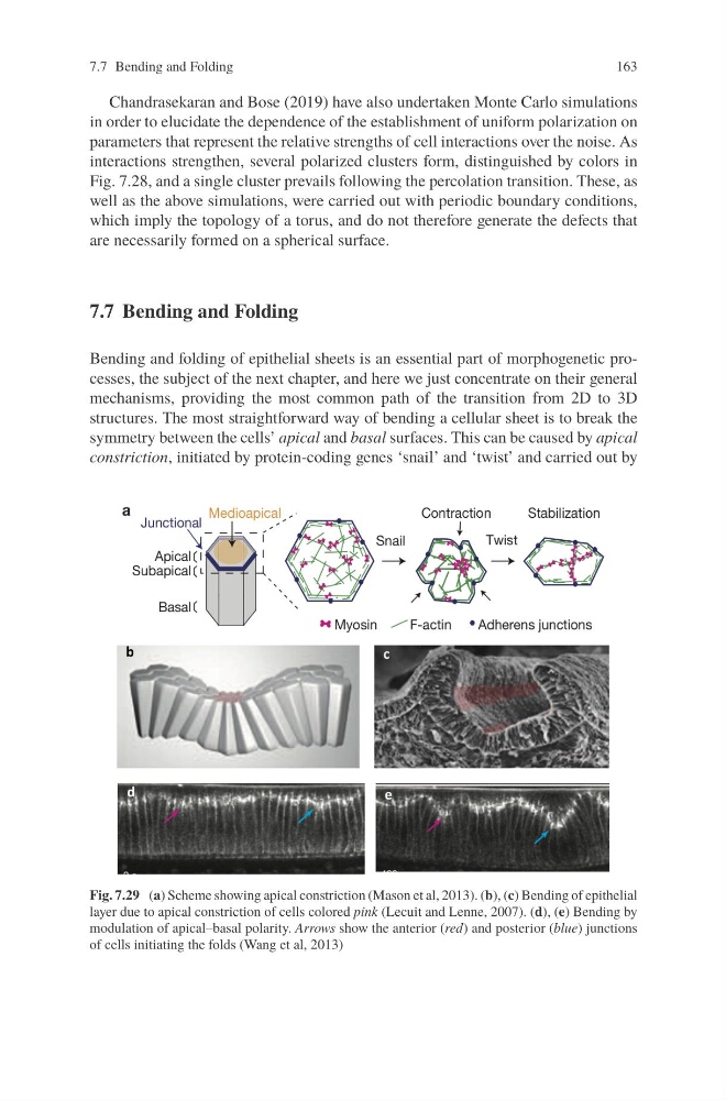
structures. The most straightforward way of bending a cellular sheet is to break the
symmetry between the cells’ apical and basal surfaces. This can be caused by apical
constriction, initiated by protein-coding genes ‘snail’ and ‘twist’ and carried out by
Fig. 7.29 (a) Scheme showing apical constriction (Mason et al, 2013). (b), (c) Bending of epithelial
layer due to apical constriction of cells colored pink (Lecuit and Lenne, 2007). (d), (e) Bending by
modulation of apical–basal polarity. Arrows show the anterior (red) and posterior (blue) junctions
of cells initiating the folds (Wang et al, 2013)
163
Chandrasekaran and Bose (2019) have also undertaken Monte Carlo simulations
in order to elucidate the dependence of the establishment of uniform polarization on
parameters that represent the relative strengths of cell interactions over the noise. As
interactions strengthen, several polarized clusters form, distinguished by colors in
Fig. 7.28, and a single cluster prevails following the percolation transition. These, as
well as the above simulations, were carried out with periodic boundary conditions,
which imply the topology of a torus, and do not therefore generate the defects that
are necessarily formed on a spherical surface.
7.7 Bending and Folding
Bending and folding of epithelial sheets is an essential part of morphogenetic processes, the subject of the next chapter, and here we just concentrate on their general
mechanisms, providing the most common path of the transition from 2D to 3D
structures. The most straightforward way of bending a cellular sheet is to break the
symmetry between the cells’ apical and basal surfaces. This can be caused by apical
constriction, initiated by protein-coding genes ‘snail’ and ‘twist’ and carried out by
Fig. 7.29 (a) Scheme showing apical constriction (Mason et al, 2013). (b), (c) Bending of epithelial
layer due to apical constriction of cells colored pink (Lecuit and Lenne, 2007). (d), (e) Bending by
modulation of apical–basal polarity. Arrows show the anterior (red) and posterior (blue) junctions
of cells initiating the folds (Wang et al, 2013)