

162
7 Live Tissues
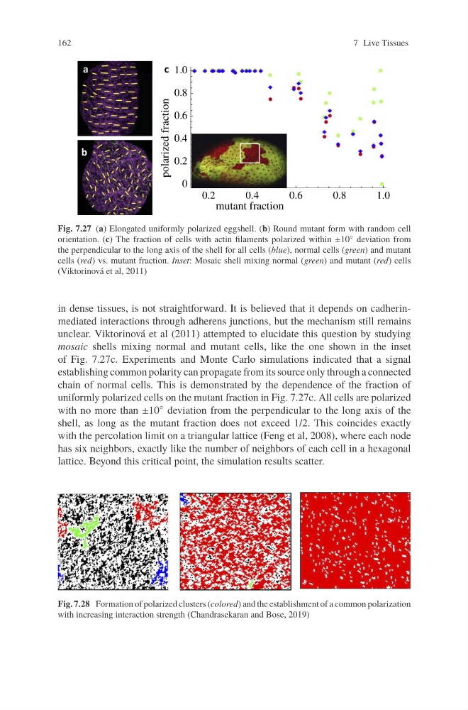
Fig. 7.27 (a) Elongated uniformly polarized eggshell. (b) Round mutant form with random cell
orientation. (c) The fraction of cells with actin filaments polarized within ±10 ◦ deviation from
the perpendicular to the long axis of the shell for all cells (blue), normal cells (green) and mutant
cells (red) vs. mutant fraction. Inset: Mosaic shell mixing normal (green) and mutant (red) cells
(Viktorinová et al, 2011)
in dense tissues, is not straightforward. It is believed that it depends on cadherinmediated interactions through adherens junctions, but the mechanism still remains
unclear. Viktorinová et al (2011) attempted to elucidate this question by studying
mosaic shells mixing normal and mutant cells, like the one shown in the inset
of Fig. 7.27c. Experiments and Monte Carlo simulations indicated that a signal
establishing common polarity can propagate from its source only through a connected
chain of normal cells. This is demonstrated by the dependence of the fraction of
uniformly polarized cells on the mutant fraction in Fig. 7.27c. All cells are polarized
with no more than ±10 ◦ deviation from the perpendicular to the long axis of the
shell, as long as the mutant fraction does not exceed 1/2. This coincides exactly
with the percolation limit on a triangular lattice (Feng et al, 2008), where each node
has six neighbors, exactly like the number of neighbors of each cell in a hexagonal
lattice. Beyond this critical point, the simulation results scatter.
Fig. 7.28 Formation of polarized clusters (colored) and the establishment of a common polarization
with increasing interaction strength (Chandrasekaran and Bose, 2019)
7 Live Tissues
Fig. 7.27 (a) Elongated uniformly polarized eggshell. (b) Round mutant form with random cell
orientation. (c) The fraction of cells with actin filaments polarized within ±10 ◦ deviation from
the perpendicular to the long axis of the shell for all cells (blue), normal cells (green) and mutant
cells (red) vs. mutant fraction. Inset: Mosaic shell mixing normal (green) and mutant (red) cells
(Viktorinová et al, 2011)
in dense tissues, is not straightforward. It is believed that it depends on cadherinmediated interactions through adherens junctions, but the mechanism still remains
unclear. Viktorinová et al (2011) attempted to elucidate this question by studying
mosaic shells mixing normal and mutant cells, like the one shown in the inset
of Fig. 7.27c. Experiments and Monte Carlo simulations indicated that a signal
establishing common polarity can propagate from its source only through a connected
chain of normal cells. This is demonstrated by the dependence of the fraction of
uniformly polarized cells on the mutant fraction in Fig. 7.27c. All cells are polarized
with no more than ±10 ◦ deviation from the perpendicular to the long axis of the
shell, as long as the mutant fraction does not exceed 1/2. This coincides exactly
with the percolation limit on a triangular lattice (Feng et al, 2008), where each node
has six neighbors, exactly like the number of neighbors of each cell in a hexagonal
lattice. Beyond this critical point, the simulation results scatter.
Fig. 7.28 Formation of polarized clusters (colored) and the establishment of a common polarization
with increasing interaction strength (Chandrasekaran and Bose, 2019)