

160
7 Live Tissues
carcinomas adopt mesenchymal features while still retaining common characteristics
of epithelial cells (Christiansen and Rajasekaran, 2006).
Moreover, cancer cells are capable to energize epithelial layers as they proliferate
and aggregate. Chen et al (2018) have found that the invasion of a small fraction of
motile cancer cells induces turbulent cooperative motion, which intensifies with the
increasing size of gradually aggregating cancer clusters and involves healthy cells
through the disruption of cell–cell junctions (Fig. 7.24).
7.6 Polarization and Defects
In living tissues, polarization is expressed on a microscopic rather than a molecular
scale and is evident in the shape of cells. Polarization can be related to the direction of
stress bundles, which often, but by no means always, coincides with both the longer
axis of the cell and the direction of motion. It may also be defined by the gradient of
some chemical cues. A change of polarization in an epithelial layer from tangential
to normal causes a conspicuous bending effect. Coupling of the polarization rotation
to elasticity can give rise to symmetry-breaking instabilities that are apt to play a
role in morphogenetic processes, as proposed by Belintsev et al (1987).
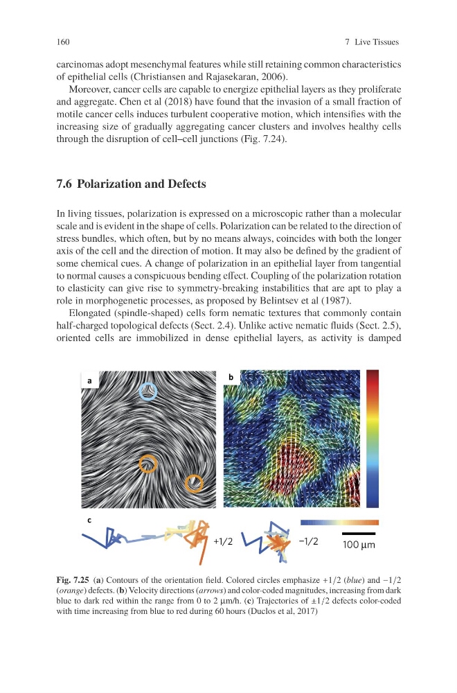
Elongated (spindle-shaped) cells form nematic textures that commonly contain
half-charged topological defects (Sect. 2.4). Unlike active nematic fluids (Sect. 2.5),
oriented cells are immobilized in dense epithelial layers, as activity is damped
Fig. 7.25 (a) Contours of the orientation field. Colored circles emphasize +1/2 (blue) and −1/2
(orange) defects. (b) Velocity directions (arrows) and color-coded magnitudes, increasing from dark
blue to dark red within the range from 0 to 2 μm/h. (c) Trajectories of ±1/2 defects color-coded
with time increasing from blue to red during 60 hours (Duclos et al, 2017)
7 Live Tissues
carcinomas adopt mesenchymal features while still retaining common characteristics
of epithelial cells (Christiansen and Rajasekaran, 2006).
Moreover, cancer cells are capable to energize epithelial layers as they proliferate
and aggregate. Chen et al (2018) have found that the invasion of a small fraction of
motile cancer cells induces turbulent cooperative motion, which intensifies with the
increasing size of gradually aggregating cancer clusters and involves healthy cells
through the disruption of cell–cell junctions (Fig. 7.24).
7.6 Polarization and Defects
In living tissues, polarization is expressed on a microscopic rather than a molecular
scale and is evident in the shape of cells. Polarization can be related to the direction of
stress bundles, which often, but by no means always, coincides with both the longer
axis of the cell and the direction of motion. It may also be defined by the gradient of
some chemical cues. A change of polarization in an epithelial layer from tangential
to normal causes a conspicuous bending effect. Coupling of the polarization rotation
to elasticity can give rise to symmetry-breaking instabilities that are apt to play a
role in morphogenetic processes, as proposed by Belintsev et al (1987).
Elongated (spindle-shaped) cells form nematic textures that commonly contain
half-charged topological defects (Sect. 2.4). Unlike active nematic fluids (Sect. 2.5),
oriented cells are immobilized in dense epithelial layers, as activity is damped
Fig. 7.25 (a) Contours of the orientation field. Colored circles emphasize +1/2 (blue) and −1/2
(orange) defects. (b) Velocity directions (arrows) and color-coded magnitudes, increasing from dark
blue to dark red within the range from 0 to 2 μm/h. (c) Trajectories of ±1/2 defects color-coded
with time increasing from blue to red during 60 hours (Duclos et al, 2017)