

7.5 Tumor Spreading
159
Fig. 7.23 Scheme of tumor spreading through repeated EMT and MET. Cancerous epithelial cells
proliferate and can undergo partial or full EMT to form heterogeneous tumors (colored) within an
otherwise non-cancerous tissue (grey). Tumors also recruit non-cancerous fibroblasts (orange) to
facilitate growth and invasion. Migratory cancer cells escape the tissue via individual or collective
migration and enter the circulatory system (red) as individual cells or as clusters, depending
on their state, distinguished by color changing from yellow (epithelial) to green (mesenchymal).
Metastatic colonization of secondary sites requires re-epithelialization of the tumor cells through
MET, resulting in the formation of secondary tumors (Plygawko et al, 2020)
and metastatic colonization and outgrowth, as sketched in Fig. 7.23. The same feature
in its benign role facilitates morphogenetic changes during normal development.
However, although EMT exacerbates motility and invasiveness of many cell types,
it is not a necessary prerequisite for tumor infiltration and metastasis, as some
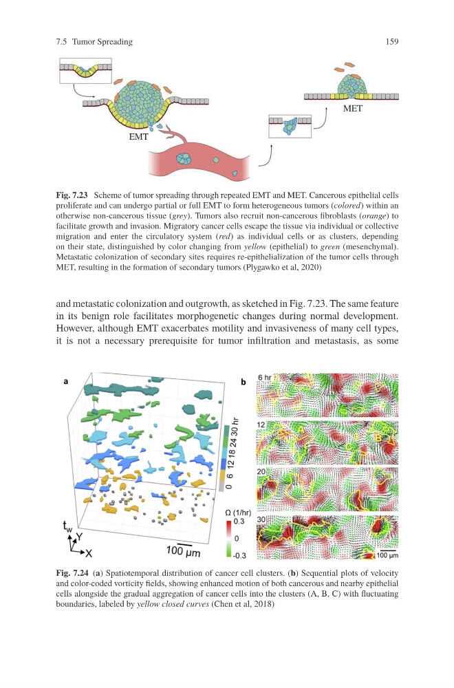
Fig. 7.24 (a) Spatiotemporal distribution of cancer cell clusters. (b) Sequential plots of velocity
and color-coded vorticity fields, showing enhanced motion of both cancerous and nearby epithelial
cells alongside the gradual aggregation of cancer cells into the clusters (A, B, C) with fluctuating
boundaries, labeled by yellow closed curves (Chen et al, 2018)
159
Fig. 7.23 Scheme of tumor spreading through repeated EMT and MET. Cancerous epithelial cells
proliferate and can undergo partial or full EMT to form heterogeneous tumors (colored) within an
otherwise non-cancerous tissue (grey). Tumors also recruit non-cancerous fibroblasts (orange) to
facilitate growth and invasion. Migratory cancer cells escape the tissue via individual or collective
migration and enter the circulatory system (red) as individual cells or as clusters, depending
on their state, distinguished by color changing from yellow (epithelial) to green (mesenchymal).
Metastatic colonization of secondary sites requires re-epithelialization of the tumor cells through
MET, resulting in the formation of secondary tumors (Plygawko et al, 2020)
and metastatic colonization and outgrowth, as sketched in Fig. 7.23. The same feature
in its benign role facilitates morphogenetic changes during normal development.
However, although EMT exacerbates motility and invasiveness of many cell types,
it is not a necessary prerequisite for tumor infiltration and metastasis, as some
Fig. 7.24 (a) Spatiotemporal distribution of cancer cell clusters. (b) Sequential plots of velocity
and color-coded vorticity fields, showing enhanced motion of both cancerous and nearby epithelial
cells alongside the gradual aggregation of cancer cells into the clusters (A, B, C) with fluctuating
boundaries, labeled by yellow closed curves (Chen et al, 2018)