

158
7 Live Tissues
7.5 Tumor Spreading
Motion and rearrangement of cells within a dense layer is essential for tissue remodeling in development; we will return to this topic in Sect. 8.6, but concentrate
here on its detrimental role in tumor spreading. In the classical view of malignant
transformation in the epithelium (Thiery, 2002), cells weaken their integrin-mediated
interactions with the extracellular matrix. Adherens junctions that are crucial for cell–
cell adhesion are subsequently lost, leading to the epithelial–mesenchymal transition
(EMT), which renders cells more motile and invasive. Single cancerous cells that
have lost contact with their surroundings can trigger metastatic spreading, and a
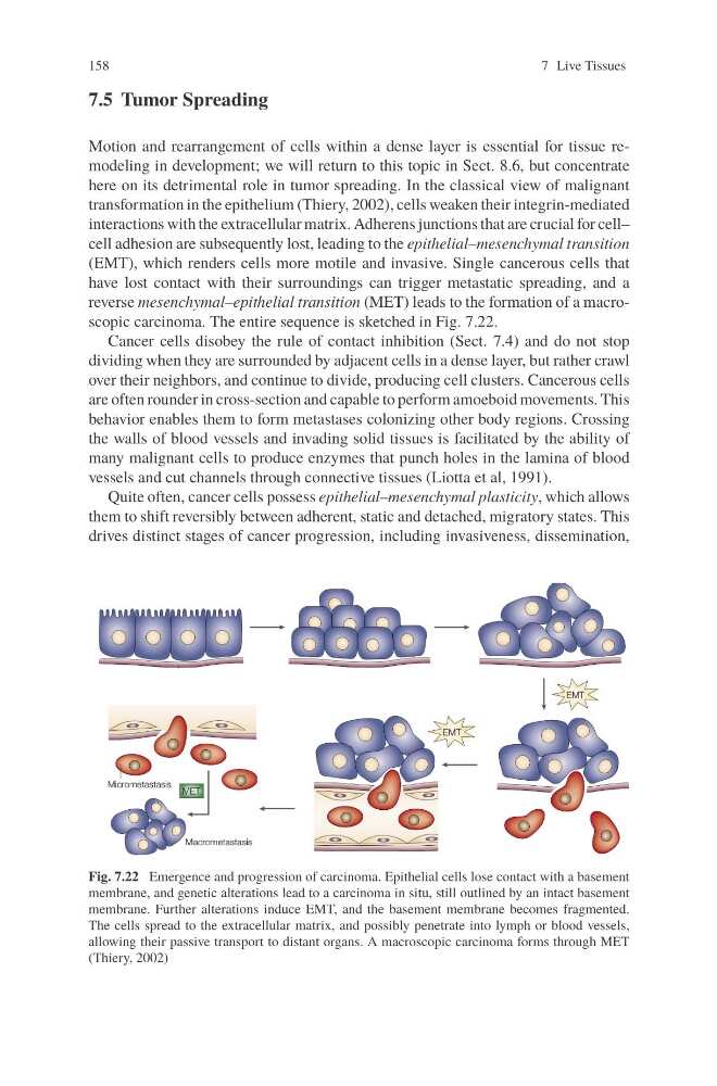
reverse mesenchymal–epithelial transition (MET) leads to the formation of a macroscopic carcinoma. The entire sequence is sketched in Fig. 7.22.
Cancer cells disobey the rule of contact inhibition (Sect. 7.4) and do not stop
dividing when they are surrounded by adjacent cells in a dense layer, but rather crawl
over their neighbors, and continue to divide, producing cell clusters. Cancerous cells
are often rounder in cross-section and capable to perform amoeboid movements. This
behavior enables them to form metastases colonizing other body regions. Crossing
the walls of blood vessels and invading solid tissues is facilitated by the ability of
many malignant cells to produce enzymes that punch holes in the lamina of blood
vessels and cut channels through connective tissues (Liotta et al, 1991).
Quite often, cancer cells possess epithelial–mesenchymal plasticity, which allows
them to shift reversibly between adherent, static and detached, migratory states. This
drives distinct stages of cancer progression, including invasiveness, dissemination,
Fig. 7.22 Emergence and progression of carcinoma. Epithelial cells lose contact with a basement
membrane, and genetic alterations lead to a carcinoma in situ, still outlined by an intact basement
membrane. Further alterations induce EMT, and the basement membrane becomes fragmented.
The cells spread to the extracellular matrix, and possibly penetrate into lymph or blood vessels,
allowing their passive transport to distant organs. A macroscopic carcinoma forms through MET
(Thiery, 2002)
7 Live Tissues
7.5 Tumor Spreading
Motion and rearrangement of cells within a dense layer is essential for tissue remodeling in development; we will return to this topic in Sect. 8.6, but concentrate
here on its detrimental role in tumor spreading. In the classical view of malignant
transformation in the epithelium (Thiery, 2002), cells weaken their integrin-mediated
interactions with the extracellular matrix. Adherens junctions that are crucial for cell–
cell adhesion are subsequently lost, leading to the epithelial–mesenchymal transition
(EMT), which renders cells more motile and invasive. Single cancerous cells that
have lost contact with their surroundings can trigger metastatic spreading, and a
reverse mesenchymal–epithelial transition (MET) leads to the formation of a macroscopic carcinoma. The entire sequence is sketched in Fig. 7.22.
Cancer cells disobey the rule of contact inhibition (Sect. 7.4) and do not stop
dividing when they are surrounded by adjacent cells in a dense layer, but rather crawl
over their neighbors, and continue to divide, producing cell clusters. Cancerous cells
are often rounder in cross-section and capable to perform amoeboid movements. This
behavior enables them to form metastases colonizing other body regions. Crossing
the walls of blood vessels and invading solid tissues is facilitated by the ability of
many malignant cells to produce enzymes that punch holes in the lamina of blood
vessels and cut channels through connective tissues (Liotta et al, 1991).
Quite often, cancer cells possess epithelial–mesenchymal plasticity, which allows
them to shift reversibly between adherent, static and detached, migratory states. This
drives distinct stages of cancer progression, including invasiveness, dissemination,
Fig. 7.22 Emergence and progression of carcinoma. Epithelial cells lose contact with a basement
membrane, and genetic alterations lead to a carcinoma in situ, still outlined by an intact basement
membrane. Further alterations induce EMT, and the basement membrane becomes fragmented.
The cells spread to the extracellular matrix, and possibly penetrate into lymph or blood vessels,
allowing their passive transport to distant organs. A macroscopic carcinoma forms through MET
(Thiery, 2002)