

7.2 Forces in Migrating Layers
149
This picture was refined with the help of modern methods of traction-force microscopy and particle image velocimetry (Brugués et al, 2014). The wound shown
in Fig. 7.10a was generated by laser ablation, and at the beginning everything proceeded along the classical scenario of cell protrusion followed by accumulation of
actin and myosin at the wound edge, as seen in Fig. 7.10b and c. Cells adjacent to
the wound became elongated, and their constricted front edges created a rosette-like
geometry. However, the velocity of each cell row around the wound exhibited a
non-monotonic time evolution: these are largest in the middle panel of the sequence
shown in Fig. 7.10d. The experimenters were surprised to observe traction forces
pointing towards the wound, colored green in Fig. 7.10e, at concave segments of the
leading edge, which showed little protrusive activity. In contrast, convex segments
showed pronounced protrusive activity and traction forces pointing away from the
wound. The difference in the rate of advance reflects the usual instability of the propagating boundary, but the reversal of traction was an unexpected effect. The traction
pattern also exhibited strong force components tangential to the wound. This pattern
is attributed to non-uniform tensions caused by the heterogeneity of the actomyosin
ring and transmitted to the underlying substrate through focal adhesions. This reveals
the important role played by the substrate. The effect is enhanced on soft substrates,
where displacements are higher, and may explain why isolated cells are slower on
soft substrates whereas wound-closure rates are not.
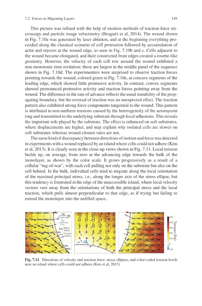
The same kind of discrepancy between directions of motion and force was detected
in experiments with a wound replaced by an island where cells could not adhere (Kim
et al, 2013). It is clearly seen in the close-up views shown in Fig. 7.11. Local tension
builds up, on average, from zero at the advancing edge towards the bulk of the
monolayer, as shown by the color scale. It grows progressively as a result of a
cellular “tug-of-war”, with each cell pulling not only on the substrate but also on the
cell behind. In the bulk, individual cells tend to migrate along the local orientation
of the maximal principal stress, i.e., along the longer axis of the stress ellipse, but
this tendency is frustrated at the edge of the unaccessible island, where local velocity
vectors veer away from the orientations of both the principal stress and the local
traction, which pulls almost perpendicular to that edge, as if trying but failing to
extend the monolayer into the unfilled space.
Fig. 7.11 Directions of velocity and traction force, stress ellipses, and color-coded tension levels
near an island where cells could not adhere (Kim et al, 2013)
149
This picture was refined with the help of modern methods of traction-force microscopy and particle image velocimetry (Brugués et al, 2014). The wound shown
in Fig. 7.10a was generated by laser ablation, and at the beginning everything proceeded along the classical scenario of cell protrusion followed by accumulation of
actin and myosin at the wound edge, as seen in Fig. 7.10b and c. Cells adjacent to
the wound became elongated, and their constricted front edges created a rosette-like
geometry. However, the velocity of each cell row around the wound exhibited a
non-monotonic time evolution: these are largest in the middle panel of the sequence
shown in Fig. 7.10d. The experimenters were surprised to observe traction forces
pointing towards the wound, colored green in Fig. 7.10e, at concave segments of the
leading edge, which showed little protrusive activity. In contrast, convex segments
showed pronounced protrusive activity and traction forces pointing away from the
wound. The difference in the rate of advance reflects the usual instability of the propagating boundary, but the reversal of traction was an unexpected effect. The traction
pattern also exhibited strong force components tangential to the wound. This pattern
is attributed to non-uniform tensions caused by the heterogeneity of the actomyosin
ring and transmitted to the underlying substrate through focal adhesions. This reveals
the important role played by the substrate. The effect is enhanced on soft substrates,
where displacements are higher, and may explain why isolated cells are slower on
soft substrates whereas wound-closure rates are not.
The same kind of discrepancy between directions of motion and force was detected
in experiments with a wound replaced by an island where cells could not adhere (Kim
et al, 2013). It is clearly seen in the close-up views shown in Fig. 7.11. Local tension
builds up, on average, from zero at the advancing edge towards the bulk of the
monolayer, as shown by the color scale. It grows progressively as a result of a
cellular “tug-of-war”, with each cell pulling not only on the substrate but also on the
cell behind. In the bulk, individual cells tend to migrate along the local orientation
of the maximal principal stress, i.e., along the longer axis of the stress ellipse, but
this tendency is frustrated at the edge of the unaccessible island, where local velocity
vectors veer away from the orientations of both the principal stress and the local
traction, which pulls almost perpendicular to that edge, as if trying but failing to
extend the monolayer into the unfilled space.
Fig. 7.11 Directions of velocity and traction force, stress ellipses, and color-coded tension levels
near an island where cells could not adhere (Kim et al, 2013)