

148
7 Live Tissues
the cause and what is the effect. As mentioned in Sect. 6.5, fingering of propagating
interfaces is a generic phenomenon. In continuous computations, it is hard to decide
whether fingers are pushed by internal dynamics, also defined as plithotaxis, from the
Greek πλ ´
ηθoς, denoting a crowd or a swarm, or pulled by kenotactic or haptotactic
action of the unoccupied area: both forces are present, and either would suffice. The
leaders may be selected and strengthened in their advance by the pushing crowd or
rise due to favorable conditions at the interface and empower followers by their pull.
Propagation of the epithelial layer is the essential part of wound healing, which,
unlike the above experiments and the modeling in Sect. 6.5 and Sect. 7.1, usually
involves closing a damaged area within the layer. It is commonly attributed to
a combination of cell crawling and “purse-string” contraction of a supracellular
actomyosin cable (Fig. 7.9), although in simple models, such as Arciero et al (2011),
wounds safely close without such an actin ring.
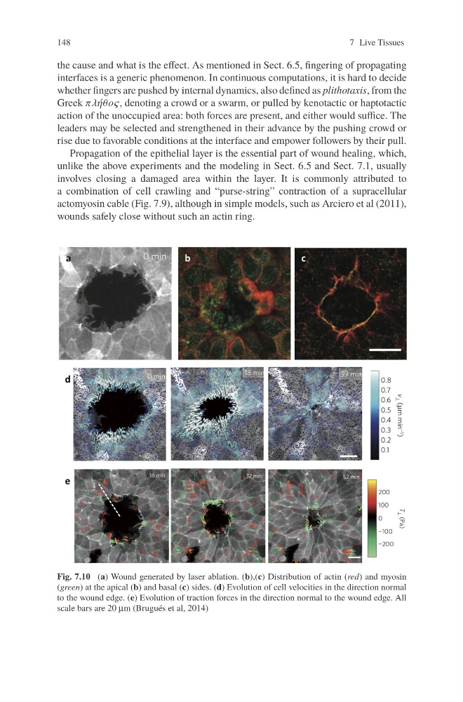
Fig. 7.10 (a) Wound generated by laser ablation. (b),(c) Distribution of actin (red) and myosin
(green) at the apical (b) and basal (c) sides. (d) Evolution of cell velocities in the direction normal
to the wound edge. (e) Evolution of traction forces in the direction normal to the wound edge. All
scale bars are 20 μm (Brugués et al, 2014)
7 Live Tissues
the cause and what is the effect. As mentioned in Sect. 6.5, fingering of propagating
interfaces is a generic phenomenon. In continuous computations, it is hard to decide
whether fingers are pushed by internal dynamics, also defined as plithotaxis, from the
Greek πλ ´
ηθoς, denoting a crowd or a swarm, or pulled by kenotactic or haptotactic
action of the unoccupied area: both forces are present, and either would suffice. The
leaders may be selected and strengthened in their advance by the pushing crowd or
rise due to favorable conditions at the interface and empower followers by their pull.
Propagation of the epithelial layer is the essential part of wound healing, which,
unlike the above experiments and the modeling in Sect. 6.5 and Sect. 7.1, usually
involves closing a damaged area within the layer. It is commonly attributed to
a combination of cell crawling and “purse-string” contraction of a supracellular
actomyosin cable (Fig. 7.9), although in simple models, such as Arciero et al (2011),
wounds safely close without such an actin ring.
Fig. 7.10 (a) Wound generated by laser ablation. (b),(c) Distribution of actin (red) and myosin
(green) at the apical (b) and basal (c) sides. (d) Evolution of cell velocities in the direction normal
to the wound edge. (e) Evolution of traction forces in the direction normal to the wound edge. All
scale bars are 20 μm (Brugués et al, 2014)