

6.6 Network Restructuring
133
be reversed as buckled filaments unbuckle once the stress is reduced, as shown by
the red curve.
Since the network is dynamic, its response to an external force depends not
only on its instantaneous strength, but on the history of forcing. In experiments by
Trepat et al (2007), a single transient stretch drove the stiffness down, whereupon it
recovered on a scale of minutes, as shown in Fig. 6.21a. The viscoelastic phase angle
δ, equal to 0 in a purely elastic medium and π/2 in a purely viscous medium, also
increases under stretch, indicating fluidization of the cytoskeleton, and gradually
recovers (Fig. 6.21b).
Wolff et al (2012), working with an in vitro model cytoskeleton, subjected it to
periodic stretching that caused the instantaneous stiffness to oscillate, as shown in the
upper panels of Fig. 6.22. Oscillations at a small strain amplitude γ (left) are regular,
but, as the amplitude increases (right), they become highly nonlinear and do not
repeat the same orbit in a chaotic fashion. In their simulations using a phenomenological extension of the wormlike chain model, they took into account a slowdown
of the long-wavelength bending undulations of the polymer backbone represented
by a stretching of the relaxation spectrum. Beyond a characteristic interaction wavelength of the order of the entanglement length, the relaxation spectrum gives rise to
a dramatic slowdown of the dynamics at long times or small frequencies, producing
the power-law strain/stress dependence that is typical for cells. At low rates, in a
quasistatic regime, it exhibits stiffening at low amplitudes, where entropic stiffening
of the polymer backbone dominates, while at high amplitudes the stiffening eventually gives way to softening, which accounts for the distinction between oscillatory
changes in the stress–strain relations. The modeling results are summarized in the
diagram of the stress–strain relations presented in the lower panel of Fig. 6.22.
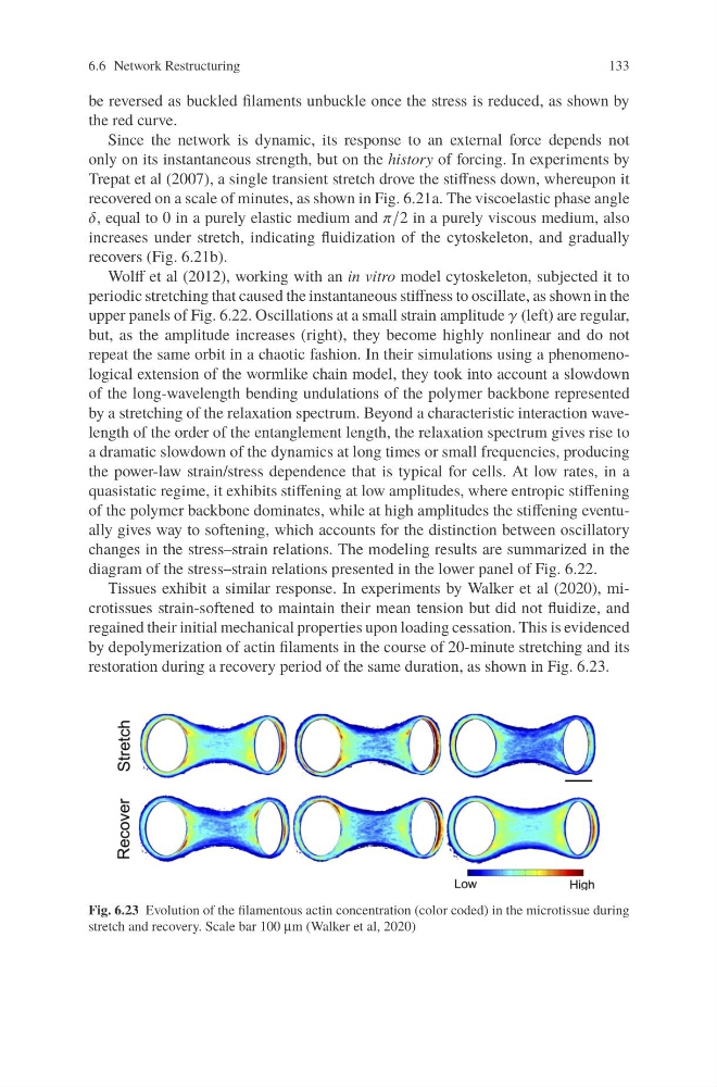
Tissues exhibit a similar response. In experiments by Walker et al (2020), microtissues strain-softened to maintain their mean tension but did not fluidize, and
regained their initial mechanical properties upon loading cessation. This is evidenced
by depolymerization of actin filaments in the course of 20-minute stretching and its
restoration during a recovery period of the same duration, as shown in Fig. 6.23.
Fig. 6.23 Evolution of the filamentous actin concentration (color coded) in the microtissue during
stretch and recovery. Scale bar 100 μm (Walker et al, 2020)
133
be reversed as buckled filaments unbuckle once the stress is reduced, as shown by
the red curve.
Since the network is dynamic, its response to an external force depends not
only on its instantaneous strength, but on the history of forcing. In experiments by
Trepat et al (2007), a single transient stretch drove the stiffness down, whereupon it
recovered on a scale of minutes, as shown in Fig. 6.21a. The viscoelastic phase angle
δ, equal to 0 in a purely elastic medium and π/2 in a purely viscous medium, also
increases under stretch, indicating fluidization of the cytoskeleton, and gradually
recovers (Fig. 6.21b).
Wolff et al (2012), working with an in vitro model cytoskeleton, subjected it to
periodic stretching that caused the instantaneous stiffness to oscillate, as shown in the
upper panels of Fig. 6.22. Oscillations at a small strain amplitude γ (left) are regular,
but, as the amplitude increases (right), they become highly nonlinear and do not
repeat the same orbit in a chaotic fashion. In their simulations using a phenomenological extension of the wormlike chain model, they took into account a slowdown
of the long-wavelength bending undulations of the polymer backbone represented
by a stretching of the relaxation spectrum. Beyond a characteristic interaction wavelength of the order of the entanglement length, the relaxation spectrum gives rise to
a dramatic slowdown of the dynamics at long times or small frequencies, producing
the power-law strain/stress dependence that is typical for cells. At low rates, in a
quasistatic regime, it exhibits stiffening at low amplitudes, where entropic stiffening
of the polymer backbone dominates, while at high amplitudes the stiffening eventually gives way to softening, which accounts for the distinction between oscillatory
changes in the stress–strain relations. The modeling results are summarized in the
diagram of the stress–strain relations presented in the lower panel of Fig. 6.22.
Tissues exhibit a similar response. In experiments by Walker et al (2020), microtissues strain-softened to maintain their mean tension but did not fluidize, and
regained their initial mechanical properties upon loading cessation. This is evidenced
by depolymerization of actin filaments in the course of 20-minute stretching and its
restoration during a recovery period of the same duration, as shown in Fig. 6.23.
Fig. 6.23 Evolution of the filamentous actin concentration (color coded) in the microtissue during
stretch and recovery. Scale bar 100 μm (Walker et al, 2020)