

6.3 Two-Phase Models
121
and cytosol are not uncommon, as reviewed by Cogan and Guy (2010) and Mogilner
and Manhart (2018). The top place in both biological detail and computational
complexity was awarded by Holmes and Edelstein-Keshet (2012) to the 3D twophase simulations by Herant and Dembo (2010). Their model viewed cytosol as
an incompressible fluid moving within the cytoskeleton as within a porous body.
The cytoskeleton was also viewed as viscous rather than elastic, and neither was its
polarization included, but its density was subject to change due to polymerization
as well as transport. The cell was kept in shape by attraction to the substrate and
specific interactions with the membrane. The latter, most ingenious part of the model
included both network–membrane repulsive stress and generation at activated portions of the three-phase contact line of a polymerization promoter diffusing to the
bulk. In this formulation, there was no need for the artificial phase field that later
came into fashion. Notwithstanding all differences between this model and the later
3D model by Tjhung et al (2015), the shapes it produced, some of which are shown
in Fig. 6.8, are rather similar. This is quite understandable. Both models contain a
number of parameters which can be adjusted to make the result look like experimentally observed crawling cells. Recall the similar convergence of 2D single-phase
models in the preceding section.
The cytoskeleton was assumed to behave on the time scale of the cell motion as
a viscous fluid in this and all the above-mentioned models, so not all ingredients
of the active gel model have been tested here. Joanny et al (2007) extended this
model to include both a viscoelastic gel and viscous fluid cytosol, as well as diffusion
equations for whatever chemical species should be essential for a particular problem.
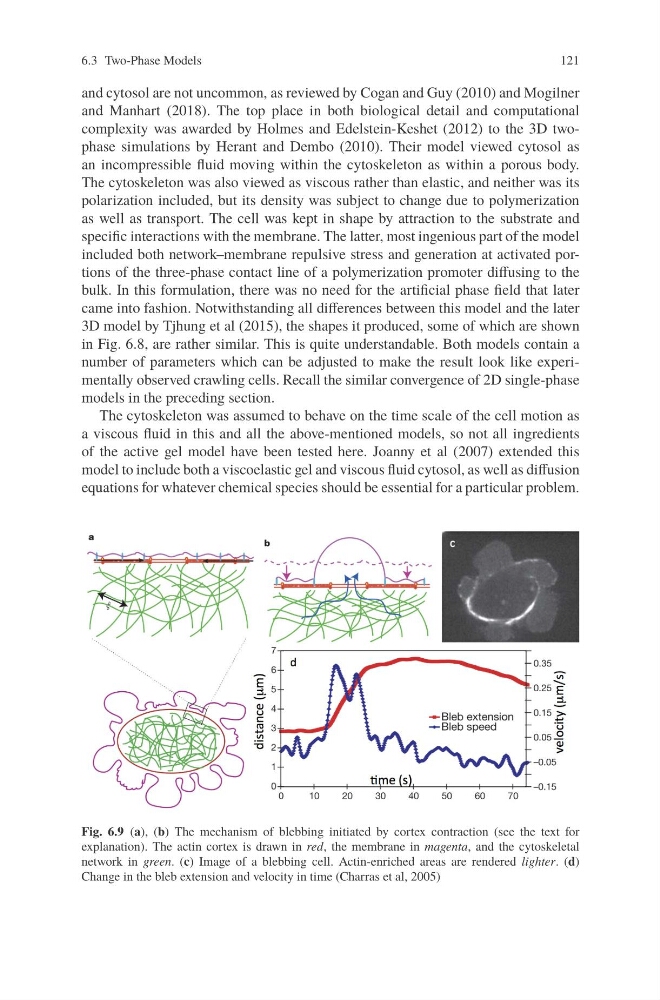
Fig. 6.9 (a), (b) The mechanism of blebbing initiated by cortex contraction (see the text for
explanation). The actin cortex is drawn in red, the membrane in magenta, and the cytoskeletal
network in green. (c) Image of a blebbing cell. Actin-enriched areas are rendered lighter. (d)
Change in the bleb extension and velocity in time (Charras et al, 2005)
121
and cytosol are not uncommon, as reviewed by Cogan and Guy (2010) and Mogilner
and Manhart (2018). The top place in both biological detail and computational
complexity was awarded by Holmes and Edelstein-Keshet (2012) to the 3D twophase simulations by Herant and Dembo (2010). Their model viewed cytosol as
an incompressible fluid moving within the cytoskeleton as within a porous body.
The cytoskeleton was also viewed as viscous rather than elastic, and neither was its
polarization included, but its density was subject to change due to polymerization
as well as transport. The cell was kept in shape by attraction to the substrate and
specific interactions with the membrane. The latter, most ingenious part of the model
included both network–membrane repulsive stress and generation at activated portions of the three-phase contact line of a polymerization promoter diffusing to the
bulk. In this formulation, there was no need for the artificial phase field that later
came into fashion. Notwithstanding all differences between this model and the later
3D model by Tjhung et al (2015), the shapes it produced, some of which are shown
in Fig. 6.8, are rather similar. This is quite understandable. Both models contain a
number of parameters which can be adjusted to make the result look like experimentally observed crawling cells. Recall the similar convergence of 2D single-phase
models in the preceding section.
The cytoskeleton was assumed to behave on the time scale of the cell motion as
a viscous fluid in this and all the above-mentioned models, so not all ingredients
of the active gel model have been tested here. Joanny et al (2007) extended this
model to include both a viscoelastic gel and viscous fluid cytosol, as well as diffusion
equations for whatever chemical species should be essential for a particular problem.
Fig. 6.9 (a), (b) The mechanism of blebbing initiated by cortex contraction (see the text for
explanation). The actin cortex is drawn in red, the membrane in magenta, and the cytoskeletal
network in green. (c) Image of a blebbing cell. Actin-enriched areas are rendered lighter. (d)
Change in the bleb extension and velocity in time (Charras et al, 2005)