

120
6 Active Gels
The equations of a viscous polar gel with boundaries imitated by the phase field
have become the prevailing choice in the single-phase continuous model. Marth et
al (2015) also included the surface tension of a cell membrane, admitting, however,
that it was of less relevance for the motility. One of the simulated sequences is shown
in Fig. 6.6. We can discern in the upper row a splay instability accompanying the
onset of motion, but it is in a direction perpendicular to the one in Fig. 6.3.
Tjhung et al (2015) extended to 3D this, as they call it, “minimal” model that
includes all components of the active gel theory except mechanical elasticity, with the
boundaries still delineated by the phase field. Several shapes observed in experiments
and approximated by the fitting parameters of the model are shown in Fig. 6.7.
6.3 Two-Phase Models
While concentrating on the cytoskeleton responsible for the cell’s mechanical
strength in the preceding chapter, we have so far passed over its liquid component – cytosol. Two-phase models including viscous motion of both cytoskeleton
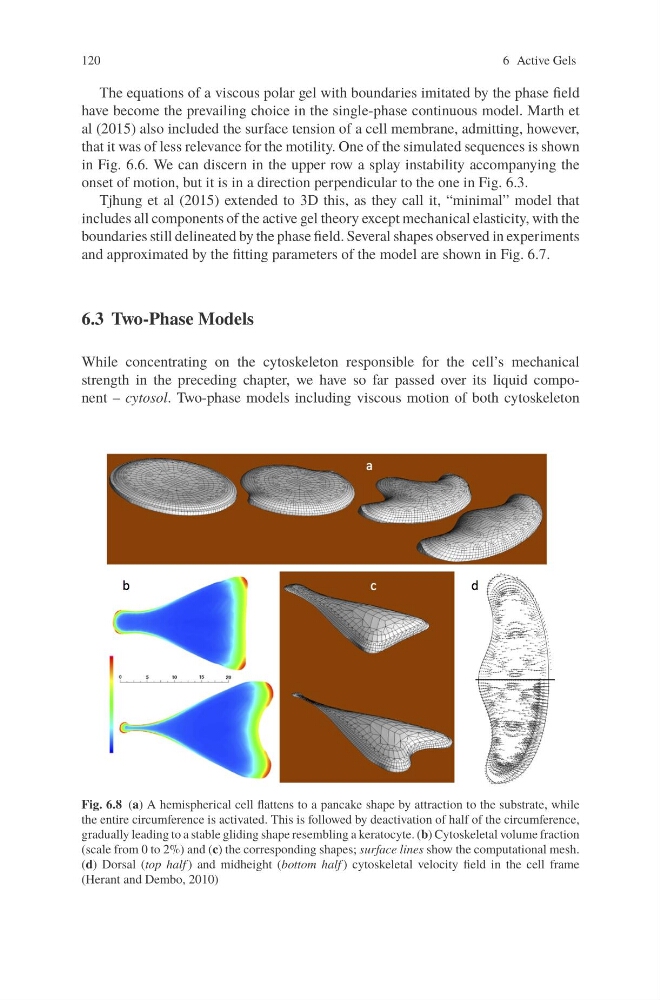
Fig. 6.8 (a) A hemispherical cell flattens to a pancake shape by attraction to the substrate, while
the entire circumference is activated. This is followed by deactivation of half of the circumference,
gradually leading to a stable gliding shape resembling a keratocyte. (b) Cytoskeletal volume fraction
(scale from 0 to 2%) and (c) the corresponding shapes; surface lines show the computational mesh.
(d) Dorsal (top half ) and midheight (bottom half ) cytoskeletal velocity field in the cell frame
(Herant and Dembo, 2010)
6 Active Gels
The equations of a viscous polar gel with boundaries imitated by the phase field
have become the prevailing choice in the single-phase continuous model. Marth et
al (2015) also included the surface tension of a cell membrane, admitting, however,
that it was of less relevance for the motility. One of the simulated sequences is shown
in Fig. 6.6. We can discern in the upper row a splay instability accompanying the
onset of motion, but it is in a direction perpendicular to the one in Fig. 6.3.
Tjhung et al (2015) extended to 3D this, as they call it, “minimal” model that
includes all components of the active gel theory except mechanical elasticity, with the
boundaries still delineated by the phase field. Several shapes observed in experiments
and approximated by the fitting parameters of the model are shown in Fig. 6.7.
6.3 Two-Phase Models
While concentrating on the cytoskeleton responsible for the cell’s mechanical
strength in the preceding chapter, we have so far passed over its liquid component – cytosol. Two-phase models including viscous motion of both cytoskeleton
Fig. 6.8 (a) A hemispherical cell flattens to a pancake shape by attraction to the substrate, while
the entire circumference is activated. This is followed by deactivation of half of the circumference,
gradually leading to a stable gliding shape resembling a keratocyte. (b) Cytoskeletal volume fraction
(scale from 0 to 2%) and (c) the corresponding shapes; surface lines show the computational mesh.
(d) Dorsal (top half ) and midheight (bottom half ) cytoskeletal velocity field in the cell frame
(Herant and Dembo, 2010)