

122
6 Active Gels
No attempt to solve this formidable system in a realistic setting has been undertaken
in this paper, which was apparently meant as an inspiration for its followers. The
antecedent of this approach lies far from biophysics. This was the poroelastic theory
originating in the studies of fluid-saturated porous soils (Biot, 1941; Coussy, 2004).
It was not mentioned by Joanny et al, but cited and applied to the model motion of
blebbing cells in the earlier paper by Charras et al (2005).
The theoretical arguments were only qualitative in this paper, but they emphasized
the elastic effect not accounted for in other models: compression of the cytosol by
the contractile cytoskeletal network. This mechanism is illustrated in Fig. 6.9a,
where part of a blebbing cell adjacent to the membrane is enlarged in the upper
panel. A local myosin-driven contraction in the cortex neighborhood (black arrows)
leads to shortening of the cortical periphery and therefore to compression of the
porous cytoskeletal network that fills the cell. The compression creates a hydrostatic
pressure, which drives the cytosol (blue arrows in Fig. 6.9b). If there is a local
defect in the attachment of the membrane to the cytoskeleton (the dashed mauve line
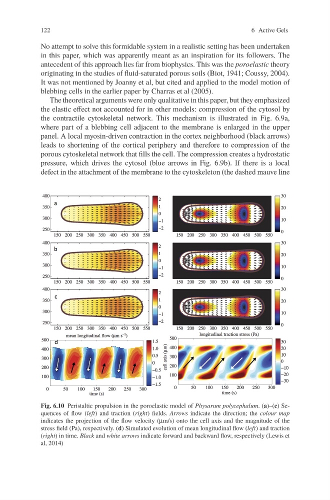
Fig. 6.10 Peristaltic propulsion in the poroelastic model of Physarum polycephalum. (a)–(c) Sequences of flow (left) and traction (right) fields. Arrows indicate the direction; the colour map
indicates the projection of the flow velocity (μm/s) onto the cell axis and the magnitude of the
stress field (Pa), respectively. (d) Simulated evolution of mean longitudinal flow (left) and traction
(right) in time. Black and white arrows indicate forward and backward flow, respectively (Lewis et
al, 2014)
6 Active Gels
No attempt to solve this formidable system in a realistic setting has been undertaken
in this paper, which was apparently meant as an inspiration for its followers. The
antecedent of this approach lies far from biophysics. This was the poroelastic theory
originating in the studies of fluid-saturated porous soils (Biot, 1941; Coussy, 2004).
It was not mentioned by Joanny et al, but cited and applied to the model motion of
blebbing cells in the earlier paper by Charras et al (2005).
The theoretical arguments were only qualitative in this paper, but they emphasized
the elastic effect not accounted for in other models: compression of the cytosol by
the contractile cytoskeletal network. This mechanism is illustrated in Fig. 6.9a,
where part of a blebbing cell adjacent to the membrane is enlarged in the upper
panel. A local myosin-driven contraction in the cortex neighborhood (black arrows)
leads to shortening of the cortical periphery and therefore to compression of the
porous cytoskeletal network that fills the cell. The compression creates a hydrostatic
pressure, which drives the cytosol (blue arrows in Fig. 6.9b). If there is a local
defect in the attachment of the membrane to the cytoskeleton (the dashed mauve line
Fig. 6.10 Peristaltic propulsion in the poroelastic model of Physarum polycephalum. (a)–(c) Sequences of flow (left) and traction (right) fields. Arrows indicate the direction; the colour map
indicates the projection of the flow velocity (μm/s) onto the cell axis and the magnitude of the
stress field (Pa), respectively. (d) Simulated evolution of mean longitudinal flow (left) and traction
(right) in time. Black and white arrows indicate forward and backward flow, respectively (Lewis et
al, 2014)